
Page 10 - Read Online
P. 10
Shetty. Plast Aesthet Res 2022;9:47 https://dx.doi.org/10.20517/2347-9264.2022.41 Page 5 of 9
Table 1. MR lymphangiography sequence parameters at 1.5 Tesla
Coronal T2-weighted single-shot Coronal heavily T2-weighted Coronal T1-weighted Dixon 3D
fast spin-echo fast spin-echo spoiled gradient echo
TR/TE (ms) 1000/100 2300/800 6.79/2.39
Flip angle (°) 150 110 10
Number of slices per 60 192 192
station
Slice thickness (mm) 4 1.5 1.5
Field of view (mm) 500 × 500 500 × 500 500 × 500
Matrix 384 × 384 512 × 512 384 × 360
Voxel size (mm) 1.3 × 1.3 × 4 1 × 1 × 1.5 1.3 × 1.3 × 1.5
Acquisition time (s) 60 250 100
Role Overview of water and fat Assessment of fluid accumulation Lymphatic and venous assessment
accumulation
Table 2. MR lymphangiography protocol at 1.5 Tesla
Sequence/Step Comments
Lymphatic intracutaneous injection of dilute gadolinium (6 mL gadolinium + 2 mL saline total), 1 mL per web space, via 25 gauge syringe,
followed by massage of web spaces to facilitate lymphatic uptake
Coronal T2-weighted single-shot fast-spin echo Reconstruct composed sequence of all stations
Coronal heavily T2-weighted fast-spin echo Reconstruct composed sequence of all stations and maximum intensity projection;
can perform in the time interval between dynamic sequences below
Coronal T1-weighted DIXON 3D spoiled gradient echo; Reconstruct water-only and fat-only images
image every 10 minutes (0, 10, 20, 30)
Intravenous injection of gadolinium contrast (weight-based dose) for venography
Coronal T1-weighted DIXON 3D spoiled gradient echo Reconstruct water-only and fat-only images; 120-second delay after contrast
venogram injection to ensure uniform venous enhancement
Coronal T2-single shot fast spin echo (large field of view Upper extremity exam only, to quantify fat accumulation compared to unaffected
with both arms) extremity
Two stations are obtained for the upper extremity using two phased array surface coils. Three stations are obtained for the lower extremities (to
include the pelvis), using a phased array surface coil over the pelvis and a peripheral angiography coil over both lower extremities.
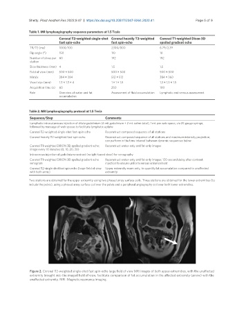
Figure 2. Coronal T2-weighted single-shot fast spin-echo large field of view MRI images of both upper extremities, with the unaffected
extremity brought into the imaged field of view, facilitate comparison of fat accumulation in the affected extremity (arrow) with the
unaffected extremity. MRI: Magnetic resonance imaging.