
Page 52 - Read Online
P. 52
Page 6 of 10 Othman et al. Plast Aesthet Res 2022;9:29 https://dx.doi.org/10.20517/2347-9264.2021.135
modality [41-43] .
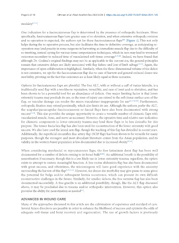
One indication for a fasciocutaneous flap is determined by the presence of orthopedic hardware. More
specifically, fasciocutaenous flaps have greater ease of re-elevation, and when extensive orthopedic revision
[44]
and re-operation is expected, the authors opt for these fasciocutaneous flaps [Figure 1]. This not only
helps during the re-operative process, but also facilitates the time to definitive coverage, as anticipating re-
operation may lend paucity in some surgeons in harvesting an immediate muscle flap due to the difficulty of
re-insetting, instead opting for various tissue temporization techniques, which in turn may lead to worsened
outcomes secondary to reduced time of vascularized soft-tissue coverage [5,30,44] . Indeed, we have found that
although Dr. Godina’s original findings may not be as applicable to the current era, the general principles
remain that extensive delays are likely associated with flap failure and loss of limb salvage [18,20] . Again, the
importance of open collaboration is highlighted. Similarly, when the three-dimensional volume of the defect
is not extensive, we opt for the fasciocutaneous flap due to ease of harvest and general reduced donor site
morbidity, pivoting on the fact that outcomes are at least likely equal in these scenarios.
Options for fascicutaneous flaps are plentiful. The free ALT, with or without a cuff of vastus lateralis, is a
traditionally used flap with a workhorse reputation, versatility, and ease of inset and re-elevation, and has
been shown to be a powerful tool for an abundance of defects. One major limiting factor is that lower
extremity trauma may preclude its use, as the zone of injury can extend to the soft-tissue surface area of the
flap, or vascular damage can render the micro-vasculature inappropriate for use [35-37,45-47] . Furthermore,
orthopedic fixation may extend proximally, which also limits its use. Although the authors prefer the ALT,
the scapular/parascapular fasciocutaneous or fascial flaps have also been documented with strong
success [45,48] . This also provides the unique opportunity to create a versatile number of chimeric flaps with
vascularized muscle, bone, and nerve as necessary. However, the operative time and relative rare indication
for chimeric components in lower extremity trauma may lend these flaps to be less desirable for this
purpose. The tensor fascia lata flap has also been used for reconstruction of the lower extremity with good
success. We also have used the lateral arm flap, though the teaching of this flap has dwindled in recent years.
Additionally, the superficial circumflex iliac artery flap (SCIP flap) has been shown to be versatile for many
purposes, though the strongest and most abundant literature comes from the Asian population, and its
validity in the western-based population is less documented due to increased obesity [49,50] .
When considering myofascial or myocutaneous flaps, the free latissimus dorsi flap has been well
documented for a number of defects owning to its broad bulk [44,45] . An additional benefit is the possibility of
neurotization if necessary, though this is a less likely use in lower extremity trauma; regardless, the option
exists to attempt to restore meaningful function. A free rectus abdominis flap has also been documented
with great success, and oftentimes, the microsurgeon will have good experience with the anatomy
surrounding the harvest of this flap [5,45,51,52] . However, the donor site morbidity may give pause to some given
the potential for bulge and/or subsequent hernia occurrence, which can present its own difficult
reconstructive challenges in the future. Similarly, for smaller defects, the free serratus flap has also been
documented successfully. A free gracilis is an additional possibility, though, like the ALT flap discussed
above, it may be precluded due to trauma and/or orthopedic intervention. However, this option also
provides the ability for neurotization as needed .
[51]
ADVANCES IN WOUND CARE
Many of the approaches discussed in this article are the culmination of experience and standard of care.
Several future directions are possible in order to enhance the likelihood of success and optimize the odds of
adequate soft-tissue and bony recovery and regeneration. The use of growth factors is previously