
Page 40 - Read Online
P. 40
[33]
as seen with a 6-month follow up. Pradhan et al. also in a
similar study found a significant difference in the postoperative
mouth opening, an insignificant difference for post surgical
morbidity and higher grades of surgical convenience in using
collagen sheet as a wound dressing material as compared to
[39]
buccal pad of fat. Reddy et al. found good results in cases of
OSMF when they impregnated dexamethasone in the collagen
graft after excision of fibrous bands.
MANIPULATION OF COLLAGEN
Though it has not been mentioned in literature, we have observed
that most surgeons find it difficult to handle the wet collagen
sheet in the oral cavity once it is taken out from its sterile packing.
Even after washing away the preservative medium by immersing
the material in sterile solution for 5-10 min, the tendency of the
collagen to coil in itself does not go away. In our opinion, it can be
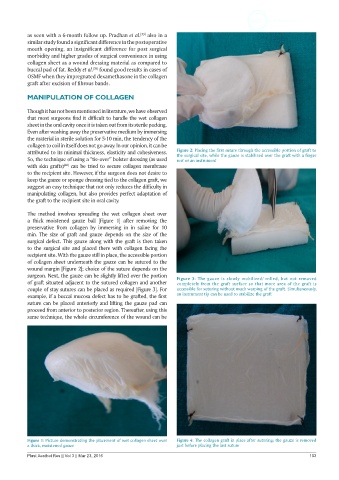
attributed to its minimal thickness, elasticity and cohesiveness. Figure 2: Placing the first suture through the accessible portion of graft to
the surgical site, while the gauze is stabilized over the graft with a finger
So, the technique of using a “tie-over” bolster dressing (as used rest or an instrument
with skin grafts) can be tried to secure collagen membrane
[40]
to the recipient site. However, if the surgeon does not desire to
keep the gauze or sponge dressing tied to the collagen graft, we
suggest an easy technique that not only reduces the difficulty in
manipulating collagen, but also provides perfect adaptation of
the graft to the recipient site in oral cavity.
The method involves spreading the wet collagen sheet over
a thick moistened gauze ball [Figure 1] after removing the
preservative from collagen by immersing in in saline for 10
min. The size of graft and gauze depends on the size of the
surgical defect. This gauze along with the graft is then taken
to the surgical site and placed there with collagen facing the
recipient site. With the gauze still in place, the accessible portion
of collagen sheet underneath the gauze can be sutured to the
wound margin [Figure 2]; choice of the suture depends on the
surgeon. Next, the gauze can be slightly lifted over the portion Figure 3: The gauze is slowly mobilized/ rolled, but not removed
of graft situated adjacent to the sutured collagen and another completely from the graft surface so that more area of the graft is
couple of stay sutures can be placed as required [Figure 3]. For accessible for suturing without much warping of the graft. Simultaneously,
example, if a buccal mucosa defect has to be grafted, the first an instrument tip can be used to stabilize the graft
suture can be placed anteriorly and lifting the gauze pad can
proceed from anterior to posterior region. Thereafter, using this
same technique, the whole circumference of the wound can be
Figure 1: Picture demonstrating the placement of wet collagen sheet over Figure 4: The collagen graft in place after suturing; the gauze is removed
a thick, moistened gauze just before placing the last suture
Plast Aesthet Res || Vol 3 || Mar 23, 2016 103