
Page 66 - Read Online
P. 66
Page 4 of 10 Schopper et al. Plast Aesthet Res 2022;9:25 https://dx.doi.org/10.20517/2347-9264.2021.72
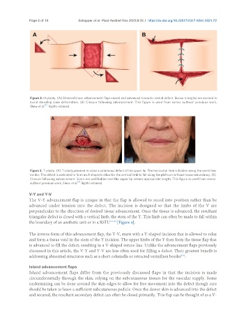
Figure 2. H-plasty. (A) Bilateral linear advancement flaps raised and advanced towards central defect. Burow triangles are excised to
avoid standing cone deformities. (B) Closure following advancement. This figure is used from senior authors’ previous work,
[1]
Shew et al. . Rights retained.
Figure 3. T-plasty. (A) T-plasty planned to close a cutaneous defect of the upper lip. The horizontal limb is hidden along the vermillion
border. The defect is extended to form an A shape to allow for the vertical limb to fall along the philtrum without tissue redundancy. (B)
Closure following advancement. Scars are well hidden and the upper lip retains appropriate length. This figure is used from senior
[1]
authors’ previous work, Shew et al. . Rights retained.
V-Y and Y-V
The V-Y advancement flap is unique in that the flap is allowed to recoil into position rather than be
advanced under tension into the defect. The incision is designed so that the limbs of the V are
perpendicular to the direction of desired tissue advancement. Once the tissue is advanced, the resultant
triangular defect is closed with a vertical limb, the stem of the Y. This limb can often be made to fall within
the boundary of an aesthetic unit or in a RSTL [11,12] [Figure 4].
The inverse form of this advancement flap, the Y-V, starts with a Y-shaped incision that is allowed to relax
and form a tissue void in the stem of the Y incision. The upper limbs of the Y then form the tissue flap that
is advanced to fill the defect, resulting in a V-shaped suture line. Unlike the advancement flaps previously
discussed in this article, the V-Y and Y-V are less often used for filling a defect. Their greatest benefit is
addressing abnormal structures such as a short columella or retracted vermillion border .
[13]
Island advancement flaps
Island advancement flaps differ from the previously discussed flaps in that the incision is made
circumferentially through the skin, relying on the subcutaneous tissues for the vascular supply. Some
undermining can be done around the skin edges to allow for free movement into the defect though care
should be taken to leave a sufficient subcutaneous pedicle. Once the donor skin is advanced into the defect
and secured, the resultant secondary defect can often be closed primarily. This flap can be thought of as a V-