
Page 47 - Read Online
P. 47
Rosi-Schumacher et al. Plast Aesthet Res 2022;9:11 https://dx.doi.org/10.20517/2347-9264.2021.64 Page 3 of 8
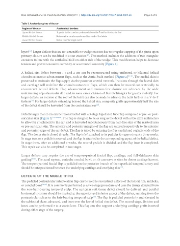
Table 1. Anatomic regions of the ear
Region of the ear Anatomical borders
Upper third of the ear Superior to the concha cymba and above the Frankfort horizontal line
Middle third of the ear Between the concha cymba and the start of the lobule
Lower third of the ear Below the intertragal notch
layers . Larger defects that are not amenable to wedge excision due to irregular cupping of the pinna upon
[16]
primary closure can be modified to a star excision . This method includes the addition of two triangular
[17]
excisions in line with the antihelical fold on either side of the wedge. This modification helps to decrease
tension and prevent excessive convexity or accentuated concavity [Figure 1].
A helical rim defect between 1.5 and 2 cm can be reconstructed using unilateral or bilateral helical
chondrocutaneous advancement flaps, such as the Antia-Buch method [Figure 2] [18-21] . The medial skin is
preserved to maintain the flap supply via the posterior arterial network. Incisions through the lateral skin
and cartilage will mobilize the chondrocutaneous flaps, which can then be moved concentrically to
reconstruct helical defects. Flap advancement and tension-free closure are achieved by the wide
undermining of postauricular skin and, in some cases, excision of Burrow triangles for greater mobility. For
larger defects, an incision at the root of the helix can also be made to advance the helix further in a V-to-Y
[17]
fashion . For larger defects extending beyond the helical rim, composite grafts approximately half the size
[22]
of the defect should be harvested from the contralateral ear .
Defects larger than 2 cm can be reconstructed with a 3-stage bipedicled tube flap composed of pre- or post-
auricular skin [Figure 3] [17,19,23,24] . The flap is designed to be as long as the defect with a few extra millimeters
to allow for attachment to the ear, and is harvested subcutaneously from hair-free skin of the mastoid area
or pre-auricular skin. The anterior and posterior margins of the flap are sutured respectively to the anterior
and posterior edges of the ear defect. The flap is tubed by suturing the free caudal and cephalic ends of the
flap. The donor site is closed directly. The flap is left attached to its pedicles for approximately three weeks.
In stage two, one pedicle is severed, and the flap is attached to the corresponding aspect of the helical defect.
In stage three, after an additional 3 weeks, the second pedicle is divided, and the flap inset is completed.
This repair can also be completed in two stages.
Larger defects may require the use of temporoparietal fascial flap, cartilage, and full-thickness skin
grafting [25,26] . The nasal septum, auricular conchal bowl, or rib can serve as sites for donor cartilage harvest.
The temporoparietal fascial flap is pedicled on the posterior branch of the superficial temporal artery and
should be interpositioned between the underlying cartilage and overlying skin .
[25]
DEFECTS OF THE MIDDLE THIRD
The pedicled postauricular interpolation flap can be used to reconstruct defects of the helical rim, antihelix,
or conchal bowl [22,27] . It is commonly performed as a two-stage procedure and uses the tissues donated from
the non-hair-bearing temporal scalp. The auricular soft tissue defect should be defined, and parallel
horizontal incisions should be marked at the superior and inferior aspect of the defect, running from the
postauricular sulcus to the hair-bearing temporal scalp . The flap is pedicled posteriorly and elevated in
[22]
the subfascial plane, advanced, and inset over the lateral helical rim defect. The second stage, division and
inset, can be performed 3 to 4 weeks later. This flap can also support underlying cartilage grafts inserted
during either stage of the surgery.