
Page 38 - Read Online
P. 38
Page 8 of 14 Brawley et al. Plast Aesthet Res 2022;9:6 https://dx.doi.org/10.20517/2347-9264.2021.107
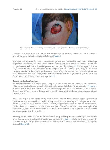
Figure 8. Defect of the occipital scalp (note: this image has been slightly altered to remove presurgical markings).
have found the posterior cervical rotation flap to have a high success rate, color/texture match, versatility,
and hairline optimization for occipital scalp lesions [Figure 9].
2
For larger defects greater than 45 cm , Orticochea flaps have been described for this location. These flaps
require vast undermining for advancement and are pedicled by bilateral superficial temporal arteries and
occipital arteries, with a three flap technique favored over a four flap technique [44,45] . Other regional flaps for
large tissue defects in this area include the temporo-parietal-occipital (Juri) flap, the trapezius
myocutaneous flap, and the latissimus dorsi myocutaneous flap. The latter two have generally fallen out of
favor due to their non-hair-bearing nature and restriction by pedicle length, especially in the era of free
flaps; however, reliable results have been reported [46-49] .
Temporoparietal scalp defects
As previously discussed, the temporoparietal scalp is the most mobile portion of the scalp with the addition
of the temporoparietal fascia, making local tissue transfer an excellent choice for reconstruction [Figure 10].
However, due to the patient’s hairline and proximity of the pinna, careful selection of local flap is needed.
Defects ranging from 2-4 cm in diameter can be closed primarily with undermining and consideration of
these structures.
The O-to-Z flap is a double rotation flap used to close a circular defect. The two opposing curvilinear
pedicles are rotated toward each other, filling the defect and creating a “Z” shaped suture line.
Buckingham et al. , based on their cadaveric research, proposed that to achieve minimal suture tension,
[50]
the lengths of each curvilinear incision should be 4 radii of the defect, with successive polar angles of 45
degrees at 2, 3, and 5 radii from the center of the defect. However, these ideal lengths can be modified with
surrounding tissue availability and laxity.
This flap can readily be used on the temporoparietal scalp, with flap design accounting for hair-bearing
areas. Camouflage with adjacent hair can be used subsequently [Figure 11]. In larger defects or areas with
less skin laxity, a skin graft can supplement the central portion after partial rotations of the flaps are
achieved [Figure 12].