
Page 74 - Read Online
P. 74
brachial plexus injuries, causes complete atrophy of target features from micro‑ to nanoscale, several surface
tissues, followed by fibrosis and fragmentation of motor modifications have been performed in order to simulate
fibers. the organized native structure of the neuronal tissue,
including micro‑ or nanogrooves to direct SC and neurite
The current “gold standard” in peripheral nerve surgeries
is an autograft, which is defined as the interposition of alignments in a mechanism also known as “conduct
[7,8]
autologous nerve segments (typically from the leg or the guidance”, micro‑pits and pillars. Microgrooves
forearm). Despite the ideal core structure provided by triggered SC alignment and migration along the pattern
[9‑11]
the autologous tissue transferred, autografts allow only direction, simulating the organized structure of the
partial functional recovery, involve double surgery and glial cells when forming the bands of Büngner. Another
cause donor tissue morbidity, calling for tissue engineered technique commonly used to recreate longitudinal
solutions to overcome these inconveniences. patterns in the conduit lumen is electrospinning, which
allows the fabrication of micro‑ or nanofibrous conduits.
A nerve guidance conduit (NGC) is a valid alternative to Nerve conduits fabricated with electrospun aligned
autograft, providing a confined environment for the entire fibers influence cell migration and nerve fiber alignment
regenerative process. NGC can be made of both natural after regeneration. Aligned micro‑ and submicro‑
[12]
[13]
[7]
and artificial materials. Its chemical and physical properties electrospun fibers were compared to a random fiber
can be optimized to achieve the best performance in configuration in an in vivo study, with the oriented
terms of tissue regeneration and inflammatory response, topography stimulating axon outgrowth and glial cell
as illustrated by several reviews. [4‑6] However, despite the migration along the direction of the fibers. Moreover,
number of proposed engineered materials, the functional variations in fiber diameter and distribution have been
recovery after conduit repair of peripheral nerve injuries shown to affect both the permeability and the porosity of
still fails where long (> 3 cm) gaps are created. the neural tube, finally influencing cell response. [4]
In the last decade, researchers have focused on different A different approach to alter the architecture of nerve
approaches to control and guide the regeneration of conduit guidance is to fill the empty tube with oriented
the injured tissue. The most promising options will be intraluminal frameworks or filaments, characterized by
discussed below, including modification of the inner lumen a larger total surface area compared to a bare conduit.
architecture, transplantation of glial/stem cells (SCs), However, these fillers may hinder the regenerative process,
inclusion of extracellular matrix (ECM) components and and it is necessary to accurately control their “packing
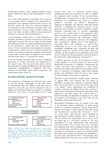
neurotrophic factors [Figure 1]. density” and distribution, which may have a large impact
on the final ability of the nerve to regenerate.
INTRALUMINAL ARCHITECTURE
Thin films of polyacrylonitrile‑co‑methyl acrylate composed
The importance of designing new NGC has been raised of aligned fibers were inserted into the lumen of
in the last decade. Topography of the inner lumen polysulfone conduits and compared to randomly aligned
can dramatically affect the ability of both the nerve to fibers and smooth films in a short‑term in vivo study using
[14]
regenerate across the gap and the endogenous cells to a rat model. Nerve regeneration was accelerated in
migrate and proliferate along the structure to modulate conduits containing the aligned fibrous film, resulting in
production and release of neurotrophic factors. Using higher levels of myelination and muscle reinnervation when
compared to the other groups. This could be due to a
high directionality and alignment of endogenous SC, which
are involved in the formation of the new tissue and the
myelination of the regenerated axons.
Microchannel elongating across the length of the tube
is an alternative lumen modification to guide axonal
growth in a confined environment. Agarose multi‑channel
conduits were shown to allow axonal growth after injury,
and vascularization occurred after 10 weeks in vivo.
[15]
In a recent study, a silicon‑based conduit containing 24
micro‑fabricated parallel channels with a diameter of
130 μm allowed the regeneration of the nerve across
the injury gap in a rat model, resulting in 85% axon
myelination. It was demonstrated that innervation
[16]
was unsuccessful at the external ring of the concentric
microchannels while all the remaining channels were filled
Figure 1: Different tissue engineering approaches to improve nerve with neuronal tissue and blood vessels. When cells were
conduits for peripheral nerve regeneration. MSC: Mesenchymal
adult stem cells, ASC: Adipose‑derived adult stem cells, LM: Laminin, preloaded in microchannel conduits, the internal guides
FN: Fibronectin, ECM: Extra cellular matrix, NGF: Nerve growth also helped the seeding and increased the availability of
factor, BDNF: Brain‑derived neurotrophic factor, NTs: Neurotrophins, [17]
GDNF: Glial‑derived neurotrophic factor, FGF: Fibroblast growth factor, the cells, with enhanced outcomes. Interestingly, when
NRG‑1: Neuregulin 1 similar multichannel structures were created with fibrin,
214 Plast Aesthet Res || Vol 2 || Issue 4 || Jul 15, 2015