
Page 56 - Read Online
P. 56
when treated within 3 months following injury, carry a muscle(s) and skin territory. A specific movement will still
high risk of incurring irreversible muscle atrophy before be performed by the original muscle, without the need to
the regenerating axons can reach the motor end plates. re‑route different tendons or muscles, which might in turn
Transferring a motor nerve that is close to the motor end lose some of their original power.
plate shortens the distance for axon regeneration and
consequently the time for muscle re‑innervation. In this RADIAL NERVE DEFICITS
respect, nerve transfer promotes a functional rather than
an anatomical reconstruction. This is the main concept Indications
[15]
in nerve transfer surgery. Other equally important concepts The radial nerve can suffer from a multitude of injuries,
include the use of tension‑free sutures directly between the with humeral fracture being the most common. [29‑31] Other
donor and recipient nerves without the use of nerve grafts causes include brachial plexus injuries, neuritis, direct
to ensure that the maximal number of regenerating axons trauma and compression. Radial nerve paralysis has been
is directed toward the end organ. By working at a location commonly treated by either neurolysis, nerve graft or
distal to the zone of injury, a pristine, vascular field can be tendon transfers with successful results. Nevertheless,
[32]
used, which will not interfere with nerve regeneration. [9‑11] some authors have reported the potential impairment of
Although sensory receptors have a wider margin for pronation following the transfer of the pronator teres (PT),
recovery even many months after the injury, earlier and unnatural coordination after tendon transfer, especially
[34]
repairs clearly lead to better outcomes. [14,16] while performing a full hand grip. [33,34] In 2002, Lowe et al.
described the possibility of transferring branches of the
Postoperative rehabilitation is facilitated when a nerve median nerve to recover wrist and finger extension in
with synergistic function is chosen for re‑innervation. [8,17‑19]
To ensure a tension‑free transfer, it is essential to dissect radial nerve palsy, alone or in conjunction with tendon
the donor nerve as distal as possible and the recipient as transfers. Since then several reports have elucidated the
[35‑38]
proximal as possible. When antagonistic nerves have been technical feasibility and the possible advantages.
used, the learning process is more difficult and the patient Nerve transfers
may require additional time to understand how to activate Motor
[20]
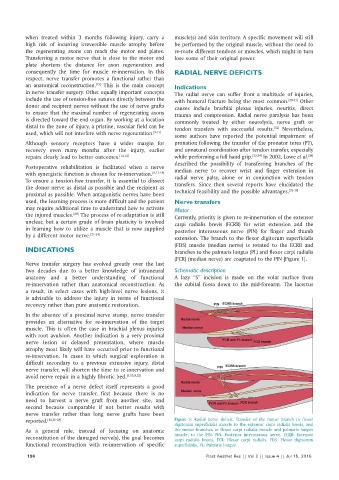
the injured muscles. The process of re‑adaptation is still Currently, priority is given to re‑innervation of the extensor
unclear, but a certain grade of brain plasticity is involved carpi radialis brevis (ECRB) for wrist extension and the
in learning how to utilize a muscle that is now supplied posterior interosseous nerve (PIN) for finger and thumb
by a different motor nerve. [21‑24] extension. The branch to the flexor digitorum superficialis
(FDS) muscle (median nerve) is rotated to the ECRB and
INDICATIONS branches to the palmaris longus (PL) and flexor carpi radialis
(FCR) (median nerve) are coaptated to the PIN [Figure 1].
Nerve transfer surgery has evolved greatly over the last
two decades due to a better knowledge of intraneural Schematic description
anatomy and a better understanding of functional A lazy “S” incision is made on the volar surface from
re‑innervation rather than anatomical reconstruction. As the cubital fossa down to the mid‑forearm. The lacertus
a result, in select cases with high‑level nerve lesions, it
is advisable to address the injury in terms of functional
recovery rather than pure anatomic restoration.
In the absence of a proximal nerve stump, nerve transfer
provides an alternative for re‑innervation of the target
muscle. This is often the case in brachial plexus injuries
with root avulsion. Another indication is a very proximal
nerve lesion or delayed presentation, where muscle
atrophy most likely will have occurred prior to functional
re‑innervation. In cases in which surgical exploration is
difficult secondary to a previous extensive injury, distal
nerve transfer, will shorten the time to re‑innervation and
avoid nerve repair in a highly fibrotic bed. [9,15,8,25]
The presence of a nerve defect itself represents a good
indication for nerve transfer, first because there is no
need to harvest a nerve graft from another site, and
second because comparable if not better results with
nerve transfer rather than long nerve grafts have been
reported. [14,26‑28] Figure 1: Radial nerve deficit. Transfer of the motor branch to flexor
digitorum superficialis muscle to the extensor carpi radialis brevis, and
As a general rule, instead of focusing on anatomic the motor branches to flexor carpi radialis muscle and palmaris longus
reconstitution of the damaged nerve(s), the goal becomes muscle, to the PIN. PIN: Posterior interosseous nerve, ECRB: Extensor
carpi radialis brevis, FCR: Flexor carpi radialis, FDS: Flexor digitorum
functional reconstruction with re‑innervation of specific superficialis, PL: Palmaris longus
196 Plast Aesthet Res || Vol 2 || Issue 4 || Jul 15, 2015