
Page 9 - Read Online
P. 9
Choudhary et al. Plast Aesthet Res 2022;9:3 https://dx.doi.org/10.20517/2347-9264.2021.68 Page 3 of 6
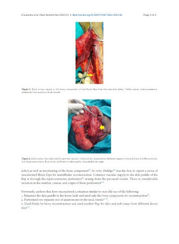
Figure 1. Black arrow: supply to the bony component of free fibula flap from the peroneal artery. Yellow arrow: septocutaneous
perforator from posterior tibial vessels.
Figure 2. Black arrow: the distal end of peroneal vessels. Yellow arrow: anastomosis between superior thyroid artery and the proximal
end of peroneal artery. Blue arrow: perforator to skin paddle, dissected to its origin.
[5]
defect as well as monitoring of the bone component . In 1989, Hidalgo was the first to report a series of
[4]
vascularized fibula flaps for mandibular reconstruction. Common vascular supply to the skin paddle of the
flap is through the septocutaneous perforators arising from the peroneal vessels. There is considerable
[4]
variation in the number, course, and origin of these perforators .
[6,7]
Previously, authors that have encountered a situation similar to ours did one of the following:
1. Returned the skin paddle to the lower limb and used only the bony component for reconstruction .
[7]
2. Performed two separate sets of anastomoses to the neck vessels [8-10] .
3. Used fibula for bony reconstruction and used another flap for skin and soft tissue from different donor
sites .
[11]