
Page 10 - Read Online
P. 10
Page 4 of 6 Choudhary et al. Plast Aesthet Res 2022;9:3 https://dx.doi.org/10.20517/2347-9264.2021.68
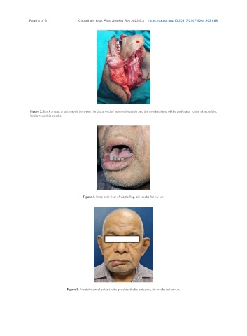
Figure 3. Black arrow: anastomosis between the distal end of peroneal vessels and the proximal end of the perforator to the skin paddle.
Red arrow: skin paddle.
Figure 4. Intra-oral view of viable flap, six weeks follow-up.
Figure 5. Frontal view of patent with good aesthetic outcome, six weeks follow-up.