
Page 23 - Read Online
P. 23
Ali et al. Plast Aesthet Res 2021;8:35 https://dx.doi.org/10.20517/2347-9264.2021.29 Page 3 of 15
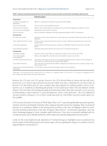
Table 1. Summary of prominent head and neck microvascular free tissue transfer reconstruction technologic advancements
Brief Description
Preoperative
Computer tomography (CT) Utilized CT angiograms for perforator mapping and MFTT design
angiography
Infrared thermography Thermal sensors used to identify perforators
Photoacoustic tomography Utilizes a near infrared pulse laser and ultrasound to provide 3D mapping of subcutaneous vessels
Color duplex ultrasonography Ultrasound used for perforator mapping
Medical modeling* Use of preoperative imaging in modeling, planning, and plating for MFTT reconstruction
Intraoperative
Microvascular couplers Alternative mode of vessel anastomosis to hand-suturing. Can be used with veins and arteries, with some
limitations
Three-dimensional (3D) exoscope 3D stereoscopic camera based viewing systems used as an alternative to operating microscopes and
surgical loupes
Fluorescent angiography Assesses arterial perfusion and venous insufficiency of the flap through the fluorescence of the skin
paddle
Osseointegrated implants* Immediate placement of implants into osseous MFTT for dental reconstruction
Postoperative
Implantable dopplers Ultrasonic probe secured to the vascular pedicle provides real-time assessment of flow
Color duplex doppler Utilizes color doppler US to identify and trace the pedicle to assess vascular flow
ultrasonography
Near-infrared spectrophotoscopy Light source emits energy at specific near-infrared wavelengths in order to measure relative changes in
concentration of oxygenated and deoxygenated hemoglobin
Laser doppler flowmetry Fiber optic probe secured to the skin paddle for the purpose of measuring microcirculatory changes via an
induced doppler shift
Digital infrared surface thermometer Thermometer monitors temperature changes in flap surface temperature
monitoring
*This topic is not discussed in this manuscript as it is covered in a separate section of this edition. CT: Computer tomography; MFTT:
microvascular free tissue transfer; 3D: three-dimensional.
between the CTA and non-CTA group. However, the CTA allowed them to choose the leg with more
perforators or to choose septocutaneous perforators which may have contributed to decreased time of
harvest. CTA also helped plan for more complex flaps that required two isolated paddles. Garvey et al.
[9]
reports a 74.1% sensitivity in identifying the presence of ALT perforators when CTA was obtained. In this
dataset, CTA was better at localizing proximal perforators than distal. They also reported a 77.5% accuracy
in determining a septocutaneous vs. musculocutaneous course of the perforator. The CTA influenced
operative decision making in 37.5% of their overall cases and over half of their cases that would require two
skin paddles.
CTA has been described for harvest of TDAP flaps. Mun et al. reported significantly decreased operative
[10]
duration and decreased length of incision when using preoperative perforator mapping. They recommend
patients to be positioned similar to the intraoperative position at the time of imaging to allow for more
[11]
accurate marking of perforators at the time of surgery. Kim and Lee recommend requesting 1 mm cuts
when obtaining imaging to better visualize small perforators. They also recommend utilizing both the
[11]
coronal and axial cuts to identify perforators off the transverse and descending thoracodorsal artery .
Lastly, for the vessel-depleted neck, dual-phase CT (delayed images to highlight venous vasculature) has
proven useful in identifying arterial and venous targets, and even guiding the surgeon to consider a local or
regional flap for reconstruction .
[12]