
Page 63 - Read Online
P. 63
Page 6 of 11 Seki et al. Plast Aesthet Res 2021;8:44 https://dx.doi.org/10.20517/2347-9264.2021.74
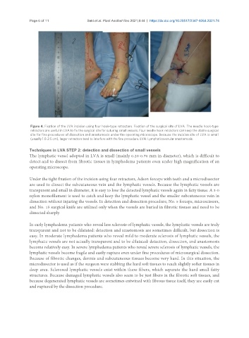
Figure 4. Fixation of the LVA incision using four hook-type retractors. Fixation of the surgical site of LVA. The needle hook-type
retractors are useful in LVA to fix the surgical site for suturing small vessels. Four needle hook retractors can keep the stable surgical
site for fine procedures of dissection and anastomosis under the operating microscope. Because the incision site of LVA is small
(usually 1.0-2.5 cm), larger retractors tend to interfere with the fine procedure. LVA: Lymphaticovenular anastomosis.
Techniques in LVA STEP 2: detection and dissection of small vessels
The lymphatic vessel adopted in LVA is small (mainly 0.20-0.70 mm in diameter), which is difficult to
detect and to dissect from fibrotic tissues in lymphedema patients even under high magnification of an
operating microscope.
Under the tight fixation of the incision using four retractors, Adson forceps with teeth and a microdissector
are used to dissect the subcutaneous vein and the lymphatic vessels. Because the lymphatic vessels are
transparent and small in diameter, it is easy to lose the detected lymphatic vessels again in fatty tissue. A 3-0
nylon monofilament is used to catch and keep the lymphatic vessel and the smaller subcutaneous vein in
dissection without injuring the vessels. In detection and dissection procedure, No. 5 forceps, microscissors,
and No. 15 surgical knife are utilized only when the vessels are buried in fibrotic tissues and need to be
dissected sharply.
In early lymphedema patients who reveal less sclerosis of lymphatic vessels, the lymphatic vessels are truly
transparent and not to be dilatated: detection and anastomosis are sometimes difficult, but dissection is
easy. In moderate lymphedema patients who reveal mild to moderate sclerosis of lymphatic vessels, the
lymphatic vessels are not actually transparent and to be dilatated: detection, dissection, and anastomosis
become relatively easy. In severe lymphedema patients who reveal severe sclerosis of lymphatic vessels, the
lymphatic vessels become fragile and easily rupture even under fine procedures of microsurgical dissection.
Because of fibrotic changes, dermis and subcutaneous tissues become very hard. In this situation, the
microdissector is used as if the surgeon were stabbing the hard soft tissues to reach slightly softer tissues in
deep area. Sclerosed lymphatic vessels exist within these fibers, which separate the hard small fatty
structures. Because damaged lymphatic vessels also seem to be just fibers in the fibrotic soft tissues, and
because degenerated lymphatic vessels are sometimes entwined with fibrous tissue itself, they are easily cut
and ruptured by the dissection procedure.