
Page 115 - Read Online
P. 115
Page 8 of 11 Seki et al. Plast Aesthet Res 2021;8:58 https://dx.doi.org/10.20517/2347-9264.2021.80
Figure 3. Definition of the incision point of the superior-edge-of-the-knee incision method. The incision point of the superior-edge-of-
the-knee incision method is 2.5 cm posterior from an intersection of transverse line drawn at the upper edge of the patella (black line)
and the longitudinal line drawn along the medial axis of the distal thigh (white line) in the supine position. At the specific point of the
incision, relatively large, less-sclerotic lymphatic vessels are always detected and utilized for an efficient functional LVA. In addition, the
subcutaneous veins with good valvar function can be detected and utilized for the recipient vein of LVA. LVA: Lymphaticovenular
anastomosis.
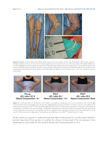
Figure 4. Continuous effect of the dynamic LVA method even under no compression. A 61-year-old woman had suffered right
International Society of Lymphology stage 2b UEL after undergoing breast cancer treatment 19 months previously. This patient is the
[20]
same patient as reported in Ref. . The preoperative right UEL index was 107.4 under sleeve compression. Three LVAs were created via
the dynamic LVA method. Soon after the surgery, lymphedema symptoms were improved. At one year, the right UEL index was reduced
to 95.7 under sleeve compression. At 18 months, she was fully released from compression therapy. At two years, the right UEL index
was 95.9 without any sleeve compression. The original source of this figure is from Ref. [20] with permission for reuse.
Further studies are required to confirm the long-term effects of functional LVA, and the authors intend to
provide long-term follow-up data to confirm the efficacy of functional LVAs and patency of the
anastomoses in a later study. We also intend to develop other functional points for LVA.