
Page 25 - Read Online
P. 25
Page 18 of 24 Reilly et al. Plast Aesthet Res 2021;8:2 I http://dx.doi.org/10.20517/2347-9264.2020.153
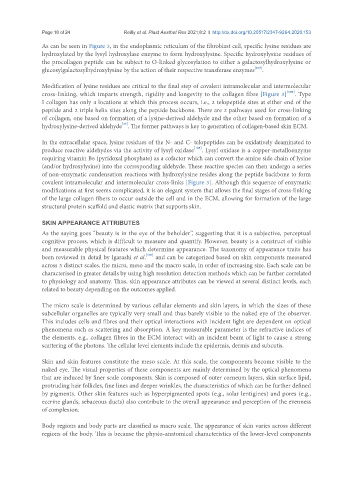
As can be seen in Figure 3, in the endoplasmic reticulum of the fibroblast cell, specific lysine residues are
hydroxylated by the lysyl hydroxylase enzyme to form hydroxylysine. Specific hydroxylysine residues of
the procollagen peptide can be subject to O-linked glycosylation to either a galactosylhydroxylysine or
glucosylgalactosylhydroxylysine by the action of their respective transferase enzymes [105] .
Modification of lysine residues are critical to the final step of covalent intramolecular and intermolecular
cross-linking, which imparts strength, rigidity and longevity to the collagen fibre [Figure 3] [106] . Type
I collagen has only 4 locations at which this process occurs, i.e., 2 telopeptide sites at either end of the
peptide and 2 triple helix sites along the peptide backbone. There are 2 pathways used for cross-linking
of collagen, one based on formation of a lysine-derived aldehyde and the other based on formation of a
[25]
hydroxylysine-derived aldehyde . The former pathways is key to generation of collagen-based skin ECM.
In the extracellular space, lysine residues of the N- and C- telopeptides can be oxidatively deaminated to
produce reactive aldehydes via the activity of lysyl oxidase [105] . Lysyl oxidase is a copper-metalloenzyme
requiring vitamin B6 (pyridoxal phosphate) as a cofactor which can convert the amine side chain of lysine
(and/or hydroxylysine) into the corresponding aldehyde. These reactive species can then undergo a series
of non-enzymatic condensation reactions with hydroxylysine resides along the peptide backbone to form
covalent intramolecular and intermolecular cross-links [Figure 3]. Although this sequence of enzymatic
modifications at first seems complicated, it is an elegant system that allows the final stages of cross-linking
of the large collagen fibers to occur outside the cell and in the ECM, allowing for formation of the large
structural protein scaffold and elastic matrix that supports skin.
SKIN APPEARANCE ATTRIBUTES
As the saying goes “beauty is in the eye of the beholder”, suggesting that it is a subjective, perceptual
cognitive process, which is difficult to measure and quantify. However, beauty is a construct of visible
and measurable physical features which determine appearance. The taxonomy of appearance traits has
been reviewed in detail by Igarashi et al. [107] and can be categorized based on skin components measured
across 3 distinct scales, the micro, meso and the macro scale, in order of increasing size. Each scale can be
characterised in greater details by using high resolution detection methods which can be further correlated
to physiology and anatomy. Thus, skin appearance attributes can be viewed at several distinct levels, each
related to beauty depending on the outcomes applied.
The micro scale is determined by various cellular elements and skin layers, in which the sizes of these
subcellular organelles are typically very small and thus barely visible to the naked eye of the observer.
This includes cells and fibres and their optical interactions with incident light are dependent on optical
phenomena such as scattering and absorption. A key measurable parameter is the refractive indices of
the elements, e.g., collagen fibres in the ECM interact with an incident beam of light to cause a strong
scattering of the photons. The cellular level elements include the epidermis, dermis and subcutis.
Skin and skin features constitute the meso scale. At this scale, the components become visible to the
naked eye. The visual properties of these components are mainly determined by the optical phenomena
that are induced by finer scale components. Skin is composed of outer corneum layers, skin surface lipid,
protruding hair follicles, fine lines and deeper wrinkles, the characteristics of which can be further defined
by pigments. Other skin features such as hyperpigmented spots (e.g., solar lentigines) and pores (e.g.,
eccrine glands, sebaceous ducts) also contribute to the overall appearance and perception of the evenness
of complexion.
Body regions and body parts are classified as macro scale. The appearance of skin varies across different
regions of the body. This is because the physio-anatomical characteristics of the lower-level components