
Page 71 - Read Online
P. 71
Göksel et al. Plast Aesthet Res 2019;6:17 I http://dx.doi.org/10.20517/2347-9264.2019.12 Page 3 of 8
Figure 1. Incision 1.2 cm in length
Figure 2. Separating the muscle fibers via blunt dissection
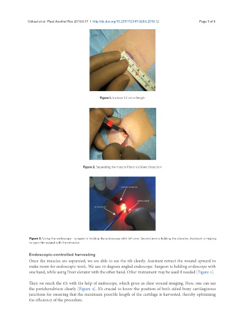
Figure 3. Using the endoscope - surgeon is holding the endoscope with left arm. Second arm is holding the elevator. Assistant is helping
to open the wound with the retractor
Endoscopic-controlled harvesting
Once the muscles are separated, we are able to see the rib clearly. Assistant retract the wound upward to
make room for endoscopic work. We use 30 degrees angled endoscope. Surgeon is holding endoscope with
one hand, while using Freer elevator with the other hand. Other instrument may be used if needed [Figure 3].
Then we reach the rib with the help of endoscope, which gives us clear wound imaging. Now, one can see
the perichondrium clearly [Figure 4]. It’s crucial to know the position of both sided bony cartilaginous
junctions for ensuring that the maximum possible length of the cartilage is harvested, thereby optimizing
the efficiency of the procedure.