
Page 84 - Read Online
P. 84
Page 4 of 6 Arkudas et al. Plast Aesthet Res 2018;5:38 I http://dx.doi.org/10.20517/2347-9264.2018.44
A B
C D
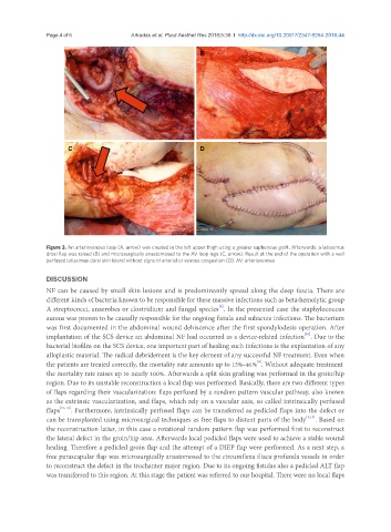
Figure 3. An arteriovenous loop (A, arrow) was created in the left upper thigh using a greater saphenous graft. Afterwards, a latissimus
drosi flap was raised (B) and microsurgically anastomosed to the AV loop legs (C, arrow). Result at the end of the operation with a well
perfused latissimus dorsi skin island without signs of arterial or venous congestion (D). AV: arteriovenous
DISCUSSION
NF can be caused by small skin lesions and is predominantly spread along the deep fascia. There are
different kinds of bacteria known to be responsible for these massive infections such as beta-hemolytic group
[9]
A streptococci, anaerobes or clostridium and fungal species . In the presented case the staphylococcus
aureus was proven to be causally responsible for the ongoing fistula and subacute infections. The bacterium
was first documented in the abdominal wound dehiscence after the first spondylodesis operation. After
[10]
implantation of the SCS device an abdominal NF had occurred as a device-related infection . Due to the
bacterial biofilm on the SCS device, one important part of healing such infections is the explantation of any
alloplastic material. The radical debridement is the key element of any successful NF treatment. Even when
[9]
the patients are treated correctly, the mortality rate amounts up to 15%-46 % . Without adequate treatment
the mortality rate raises up to nearly 100%. Afterwards a split skin grafting was performed in the groin/hip
region. Due to its unstable reconstruction a local flap was performed. Basically, there are two different types
of flaps regarding their vascularization: flaps perfused by a random pattern vascular pathway, also known
as the extrinsic vascularization, and flaps, which rely on a vascular axis, so called intrinsically perfused
flaps [11,12] . Furthermore, intrinsically perfused flaps can be transferred as pedicled flaps into the defect or
can be transplanted using microsurgical techniques as free flaps to distant parts of the body [3,13] . Based on
the reconstruction latter, in this case a rotational random pattern flap was performed first to reconstruct
the lateral defect in the groin/hip area. Afterwards local pedicled flaps were used to achieve a stable wound
healing. Therefore a pedicled groin flap and the attempt of a DIEP flap were performed. As a next step, a
free parascapular flap was microsurgically anastomosed to the circumflexa iliaca profunda vessels in order
to reconstruct the defect in the trochanter major region. Due to its ongoing fistulas also a pedicled ALT flap
was transferred to this region. At this stage the patient was referred to our hospital. There were no local flaps