
Page 83 - Read Online
P. 83
Arkudas et al. Plast Aesthet Res 2018;5:38 I http://dx.doi.org/10.20517/2347-9264.2018.44 Page 3 of 6
A
B
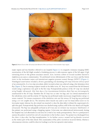
Figure 2. Before (A) and after (B) radical debridement of the scar tissue in the trochanter major region
major region and was therefore referred to our hospital [Figure 1]. An magnetic resonance imaging (MRI)
examination of the left thigh revealed a fistulating process in subcutaneous region of the left upper thigh
extending down to the gluteus maximus muscle. Also, bacteria culture of wound exudates showed a
staphylococcus aureus contamination. We performed several debridements of the scar tissue and the fistula
down to the trochanter major and intermittent negative pressure wound therapy (NPWT) [Figure 2].
Histological examination revealed a chronic granulating and ulcerating soft tissue infection without proof
of an osteomyelitis. In a two-step procedure, defect reconstruction was achieved using a free latissimus dorsi
flap [Figure 3]. Due to missing recipient vessels in the defect area, AV loop was created from the left femoral
vessels using a saphenous vein graft in the first step. Postoperatively patency of the AV loop was checked
using Doppler ultrasound. After four days a free myocutaneous latissimus dorsi flap was microsurgically
anastomosed to the AV loop. Therefore the AV loop was cut into two legs, and the arterial anastomosis of
the subclavian artery and the arterial AV loop leg was performed under microscope magnification using 8-0
suture material. The venous anastomosis of the subclavian vein and the venous AV loop leg was performed
using a 4.0 mm coupler device. The latissimus dorsi muscle was used to seal the tissue defect down to the
trochanter major whereas the skin island was inserted to close the skin defect without the requirement of a
split skin graft. Postoperatively flap perfusion was checked using capillary refill of the skin island and Doppler
ultrasound. The flap was adequately perfused at all times and no revision war necessary. Mobilization of
the patient was performed using a dangling regime. Cefuroxim was administered intravenously during
the hospital stay and afterwards in oral form for a total of six weeks postoperatively. Due to a postoperative
anemia the patient received two red cell concentrates in the further course. The patient was discharged from
the clinic 13 days after free flap transplantation. In the further course a wound healing disorder occurred
in the ventral part of the skin island. MRI examination of the pelvic showed no recurrent fistula, whereas a
subcutaneous infection could be observed leading to no further operative intervention.