
Page 76 - Read Online
P. 76
Matiasek et al. Plast Aesthet Res 2018;5:36 I http://dx.doi.org/10.20517/2347-9264.2018.50 Page 7 of 11
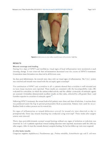
Figure 6. Ischial pressure ulcer after wound closure with posterior thigh flap
RESULTS
Wound coverage and healing
During the 6 days of NPWT and instillation, visual signs of local inflammation were monitored at each
dressing change. It was observed that inflammation decreased over the course of NPWTi treatment.
Granulation tissue formation was observed in all thirteen cases.
Six days post-debridement, the wounds were clean with no visual signs of inflammation. The V.A.C. system
was removed and wounds were rinsed with the antiseptic agent octenisept®.
The combination of NPWT and octenidine in all 13 patients showed that octenidine is well tolerated and
no toxic tissue reactions were reported. These results are consistent with the biocompatibility index (BI)
evaluated for octenidine, in which the antimicrobial activity and the cellular cytotoxicity of antiseptic agents
are assessed. Octenidine demonstrated excellent results on this index, reflected by a BI greater than 1 and
[23]
therefore superior to a number of antiseptic agents .
Following NPWTi treatment, the wound beds of all patients were clean and clear of infection. A suction drain
was positioned under the flap to prevent postoperative fluid accumulation. Patients were cared for on air-
fluidised beds to reduce pressure on the wound site.
No signs of inflammation or wound dehiscence around the wound site were observed on day 10
postoperatively. Every day, wound cleansing was conducted using octenisept®. Three weeks after surgery
sutures were removed.
Thirty days post-debridement, normal wound healing without any signs of irritation or infection was
observed. In 6 of 13 patients superficial wound healing disorders were reported, maximum until the 25th day
after surgery. After 90 days the wounds showed complete healing. No further follow-up visits were required.
In vitro tests results
Coagulase negative staphylococci, Pseudomonas spp., Proteus mirabilis, Acinetobacter spp. and E. coli were