
Page 37 - Read Online
P. 37
Polykandriotis et al. Plast Aesthet Res 2018;5:37 I http://dx.doi.org/10.20517/2347-9264.2018.52 Page 3 of 10
A B
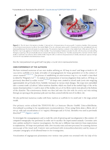
Figure 2. A: The AV-loop in the isolation chamber. V-femoral vein, A-femoral artery, G-venous graft, C-isolation chamber. After exposure
of the femoral neurovascular bundle at the medial thigh of the rat, a femoral venous graft is harvested from the contralateral side and
interposed between the femoral vessels by anastomoses. The isolation chamber is placed in the medial thigh of the rat and 300 µL of
the fibrin matrix are applied at the bottom of the chamber. The arteriovenous fistula is laid onto the clot with the artery and vein exiting
through an opening at the proximal pole and are covered with the rest of the fibrin clot. The lid is closed and the chamber with the matrix
inside is fixed onto the adductor fascia at the medial thigh; B: after an interval of approximately 6 weeks, neovascular sprouts emerge
from the AV-loop and form new fibrovascular tissue
that the interpositional vein graft itself may play a crucial role in neovascularization.
OWN EXPERIENCE OF THE AUTHORS
We have reviewed numerous of our own studies utilizing an AV-loop in small and large animals to 3D
vascularize scaffolds or to study principles of neoangiogenesis for tissue generation or in the context of
cancer research [3,7,10,11,17-22] . The process of establishing an arteriovenous loop in a rat model is described
[23]
in detail by Weigand et al. . To analyse the phenomenon of neovasculogenesis from such an AV-loop, as
previously described in earlier studies [13,14,24-26] AV-loops were created in inbred male Lewis rats weighing
approximately 250 g (Charles River, Sulzfeld, Germany) in various study designs. The loops were embedded
into a custom-made cylindrical Teflon isolation chamber, which was fixed in the medial thigh of the rat by
means of polypropylene 5-0 and in most of the studies 300 µL of the fibrin matrix were placed at the bottom
of the chamber. The arteriovenous fistula was then laid onto this clot with the artery and vein exiting
through an opening at the proximal pole and was then covered with the rest of the fibrin clot.
We also performed numerous studies with bone matrices or scaffolds in the small and in the large animal
model.
Our primary series utilized the TISSUCOL-Kit 2,0 Immuno (Baxter GmbH, Unterschleißheim,
Deutschland) according to the manufacturers recommendations. When using fibrin alone a fibrin clot of
600 µL with end concentrations of 33.7 mg/mL Fibrinogen and 25 IU/mL Thrombin was used as previously
described [13,23,27] [Figures 3 and 4].
To investigate the neoangiogenesis and to study the cycle of sprouting and vasculogenesis in this context, 3D
computed tomography was performed in order not to sacrifice the experimental animals. Corrosion casts
were antoher method to visualize vasculogenesis. For this end at different time intervals from implantation
to explantation, AV-loops were perfused with a low viscosity resin and were processed for scanning electron
microscopy of the vessel wall. Controls were performed with immunohistochemistry and by means of
computer tomography which allowed linear in vivo investigations.
Visualization of angiogenesis phenomena over various time points was attained with the help of the