
Page 39 - Read Online
P. 39
Page 6 of 9 Kovar et al. Plast Aesthet Res 2019;6:10 I http://dx.doi.org/10.20517/2347-9264.2019.09
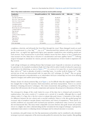
Table 3. Flap related complications compared between groups by vascular pedicle coverage
Complication Skin graft to pedicle (n = 13) Pedicle covered (n = 63) Odds ratio P value
Flap outcomes
Any flap complication 4 (31%) 19 (30.1%) 1.7 0.44
Early than 7 days take back 2 (15%) 8(12.6%) 1.9 0.46
Later than 7 days surgery 1 (7%) 9 (14.2%) 0.64 0.69
Venous thrombosis 2 (15%) 5 (7.9%) 4.8 0.11
Arterial thrombosis 0 (0%) 3 (4.7%) - -
Arterial insufficiency 0 (0%) 1 (1.6%) - -
Partial flap loss skin soft tissue fascia 1 (7%) 3 (4.7%) 3.3 0.34
Complete flap loss 1 (7%) 4 (6.3%) 1.58 0.69
Flap infection 0 (0%) 4 (6.3%) - -
Dehiscence 2 (15%) 4 (6.3%) 3.56 0.11
Contour surface irregularity 0 (0%) 0 (0%) - -
Debulking 1 (7%) 2 (3.2%) 3.2 0.35
Flap elevation 1 (7%) 9 (14.3%) 0.64 0.69
Flap removal 0 (0%) 5 (7.9%) - -
Amputation 0 (0%) 2 (3.1%) - -
Follow up average 16.2 months 22.21 months P > 0.05
compliance, elasticity, and ultimately the blood flow through the vessel. These damaged vessels are used
for the anastomosis in a free flap [15-17] . Lee et al. demonstrated that patients with serum Creatinine
[15]
greater than 1.28 mg/dL had significantly higher post-operative complication rates including, partial and
total necrosis. Flap reconstruction to previously radiated tissues has been shown to have complication
rates between 8%-39%, likely due to reduction in vascularization and mean capillary lumen [18,19] . Utilizing
surgical techniques to minimize the tension, pressure, and manipulation of these vessels is imperative for
flap survival.
Limb salvage techniques are utilizing thinner flap techniques more frequently as outcomes are proving to
be equivalent to the standard anterolateral thigh (ALT) flap with the added benefit of reduced re-operations
and revisions for debulking, as seen in Figure 1 [20,21] . In a series of 25 super-thin or supra-fascial ALT
flaps, Seth et al. had no episodes of partial or total flap losses. In another study by Hong et al. , a flap
[7]
[21]
[16]
survival rate of 98% was demonstrated with the super-thin ALT technique. Dr. Hong , like our group,
identified preoperative revascularization as an independent risk factor toward flap loss even when utilizing
supermicrosurgery and outflow preserving techniques.
Primary closure of a fasciocutaneous flap, as in Figure 2, can be difficult when the original defect’s contour
has changed due to long OR exposure, fluid resuscitation, and tissue manipulation. There is no current
literature evaluating alternative techniques. This study demonstrates skin grafting as a method for flap
closure that will minimize risk of vascular compromise and maintain the preoperative dimensions of the flap.
The retrospective design of this study limits the scope of the data that is obtained and potential for
randomization. Our series consists of 75 patients over a period of 36 months. Although this is a small
volume study of 76 flaps, this is the first series to evaluate skin graft closure. A limitation of this study is
the difference between the two cohorts and co-morbid conditions. Patients in the skin grafted group had
increased rates of peripheral vascular disease, renal disease, and history of amputations. These specific co-
morbid conditions are associated with increased post-operative complication rates; however, our study
did not demonstrate this. This study found comparable outcomes between skin graft closure and primary
closure in a higher risk cohort, demonstrating another indication for this surgical technique. A prospective,
randomized study aimed at evaluating closure techniques and the clinical outcomes is necessary.
In conclusion, skin graft closure over free flap anastomoses and pedicles may be considered a safe, alternative
technique to prevent compression of the microvascular pedicle for extremity free tissue transfers. Skin grafting