
Page 38 - Read Online
P. 38
Kovar et al. Plast Aesthet Res 2019;6:10 I http://dx.doi.org/10.20517/2347-9264.2019.09 Page 5 of 9
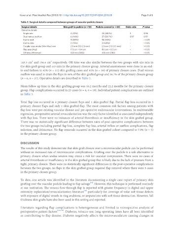
Table 2. Surgical details compared between groups of vascular pedicle closure
Surgical details Skin graft to pedicle (n = 13) Pedicle covered (n = 63) Odds ratio P value
Operative details
Single vein 4 (31%) 24 (38.1%) 1 0.96
Dual venous outflow 6 (46%) 37 (58.7%) 0.97 0.97
End-to-end 11 (85%) 58 (92%) - > 0.05
End-to-side 2 (15%) 3 (4.8%) - > 0.05
Coupler sizes mode (Min-Max) mm 2.0 mm (1.5-2.5 mm) 2.5 mm (1.5-3.5 mm) - > 0.05
Flap area (Avg) 17.3 cm × 8.4 cm 19.1 cm × 8.2 cm - > 0.05
OR time (Minimun) 420 min (340) 445 min (180) - > 0.05
2
2
145.3 cm and 156.6 cm respectively. OR time was also similar between the two groups with 420 min in
the skin graft group and 445 min in the primary closure group. Arterial anastomoses were done in an end-
to end fashion in 82% (n = 11) skin grafting cases and 92% (n = 58) of primary closure cases. Dual venous
outflow was used to drain the flaps in 46% of the skin grafted group and 58.7% of the primary closure group
(n = 6, n = 37). Operative details are described in Table 2.
Mean follow up time in the skin grafting group was 16.2 months and 22.2 months for the primary closure
group. Flap complications occurred in 23 cases (n = 4, n = 19). Individual patient complications are outlined
in Table 3.
Total flap loss occurred in 4 primary closure flaps and 1 skin grafted flap. Partial flap loss occurred in 3
primary closure flaps and only 1 skin grafted flap. The most common risk factors among patients with
flap loss were pre-existing vascular disease and pre-operative endovascular interventions. In multivariable
regression, preoperative arterial revascularization was the only factor identified as associated independently
with flap loss. There were no instances of arterial thrombosis or insufficiency in the skin grafted group.
There was no statistically significant difference between rates of post-operative complications between
the two groups including partial flap loss, complete flap loss, arterial inflow or outflow complications, flap
infection, and dehiscence. No flap removals occurred in the skin grafted cohort compared to 7.9% (n = 5)
in the primary closure group.
DISCUSSION
The results of this study demonstrate that skin graft closure over a microvascular pedicle can be performed
without an increased rate of microvascular complications. Grafting over the pedicle is a safe alternative to
primary closure when undue tension may create a risk for vascular compression. There were no cases of
arterial thrombosis or insufficiency in the skin grafted group that is likely due to the lack of pressure from a
tight, primary closure. There were no statistically significant differences in the post-operative complications
between the two groups, no flaps in the skin grafted group required flap removal where there were 5 cases
in the primary closure group.
To date, one article was identified in the literature documenting a single case report of primary skin
[11]
grafting over the vascular pedicle leading to flap salvage . However, this technique is performed routinely
at our institution. The venous flow-through flap is reported with greater frequency in digital and upper
[12]
extremity replantation/revascularization literature particularly for coverage of volar soft tissue defects
with exposure of digital vessels in ring avulsions, or amputations with soft tissue destruction. However, full
thickness skin grafts have also been used in this setting and reported.
Literature regarding flap complications is heterogeneous and limited to retrospective analysis of
perioperative patient factors [3,13,14] . Diabetes, tobacco use, long operating times have all been identified
as contributing to flap demise. Diabetes negatively affects the microvasculature causing changes in