
Page 14 - Read Online
P. 14
Tejiram et al. Plast Aesthet Res. 2025;12:9 https://dx.doi.org/10.20517/2347-9264.2024.109 Page 9 of 16
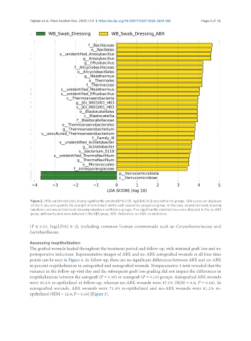
Figure 2. LEfSe enrichment plots display significantly enriched [P ≤ 0.05, log(LDA) ≥ 2] taxa within the groups. LDA scores are displayed
on the X-axis and quantify the strength of enrichment within each respective categorical group, in this case, wound bed swab dressing
takedown and wound bed swab dressing takedown antibiotics groups. Two significantly enriched taxa were detected in the no-ABX
group, and twenty-nine were detected in the ABX group. ABX: Antibiotics; no-ABX: no antibiotics.
[P ≤ 0.05, log(LDA) ≥ 2], including common human commensals such as Corynebacteriaceae and
Lactobacillaceae.
Assessing reepithelization
The grafted wounds healed throughout the treatment period and follow-up, with minimal graft loss and no
postoperative infections. Representative images of ABX and no-ABX autografted wounds at all four time
points can be seen in Figure 4. At follow-up, there are no significant differences between ABX and no-ABX
in percent reepithelization in autografted and xenografted wounds. Nonparametric t-tests revealed that the
variance in the follow-up visit day and the subsequent graft loss grading did not impact the differences in
reepithelization between the autograft (P = 0.38) or xenograft (P = 0.19) groups. Autografted ABX wounds
were 89.2% re-epithelized at follow-up, whereas no-ABX wounds were 87.1% (SEM = 9.8; P = 0.84). In
xenografted wounds, ABX wounds were 71.9% re-epithelized and no-ABX wounds were 81.2% re-
epithelized (SEM = 12.8; P = 0.49) [Figure 5].