
Page 13 - Read Online
P. 13
Page 8 of 14 Olla et al. Plast Aesthet Res 2024;11:24 https://dx.doi.org/10.20517/2347-9264.2024.30
Figure 4. Axial MRA images of the thigh for the patient in Figure 1. The perforator labeled R3 measures 1.5 mm in diameter (A). The
perforator labeled L2 measures 1.7 mm in diameter (B). (MRA = magnetic resonance angiography).
Figure 5. Volumetric assessment of right posterior thigh donor site. The estimated fat volume of a 6 × 22 cm flap on the posterior right
thigh is 499 cc.
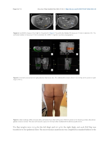
Figure 6. Initial markings of the profunda artery perforator flap in the holding area. With the patient in the standing position, the inferior
gluteal crease is marked. The ideal soft tissue for capture in the flap is then marked with the dotted purple marker.
The flap weights were 512 g for the left thigh and 507 g for the right thigh, and each PAP flap was
transferred to the ipsilateral chest. The microvascular anastomosis was completed in standard fashion to the