
Page 68 - Read Online
P. 68
Hosomi et al. Plast Aesthet Res 2023;10:60 https://dx.doi.org/10.20517/2347-9264.2023.77 Page 5 of 8
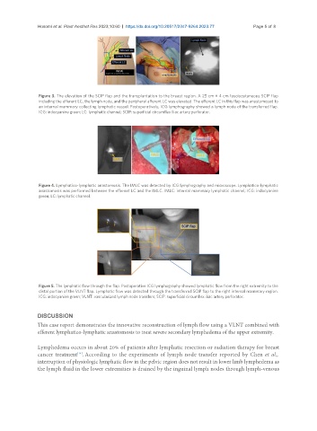
Figure 3. The elevation of the SCIP flap and the transplantation to the breast region. A 25 cm × 4 cm fasciocutaneous SCIP flap
including the efferent LC, the lymph node, and the peripheral afferent LC was elevated. The efferent LC in this flap was anastomosed to
an internal mammary collecting lymphatic vessel. Postoperatively, ICG lymphography showed a lymph node of the transferred flap.
ICG: indocyanine green; LC: lymphatic channel; SCIP: superficial circumflex iliac artery perforator.
Figure 4. Lymphatico-lymphatic anastomosis. The IMLC was detected by ICG lymphography and microscope. Lymphatico-lymphatic
anastomosis was performed between the efferent LC and the IMLC. IMLC: internal mammary lymphatic channel; ICG: indocyanine
green; LC: lymphatic channel.
Figure 5. The lymphatic flow through the flap. Postoperative ICG lymphography showed lymphatic flow from the right extremity to the
distal portion of the VLNT flap. Lymphatic flow was detected through the transferred SCIP flap to the right internal mammary region.
ICG: indocyanine green; VLNT: vascularized lymph node transfers; SCIP: superficial circumflex iliac artery perforator.
DISCUSSION
This case report demonstrates the innovative reconstruction of lymph flow using a VLNT combined with
efferent lymphatico-lymphatic anastomosis to treat severe secondary lymphedema of the upper extremity.
Lymphedema occurs in about 20% of patients after lymphatic resection or radiation therapy for breast
cancer treatment . According to the experiments of lymph node transfer reported by Chen et al.,
[14]
interruption of physiologic lymphatic flow in the pelvic region does not result in lower limb lymphedema as
the lymph fluid in the lower extremities is drained by the inguinal lymph nodes through lymph-venous