
Page 56 - Read Online
P. 56
Rajaram et al. Plast Aesthet Res. 2025;12:6 https://dx.doi.org/10.20517/2347-9264.2024.147 Page 7 of 13
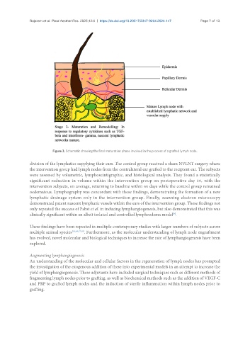
Figure 3. Schematic showing the final maturation phase involved in the process of a grafted lymph node.
division of the lymphatics supplying their ears. The control group received a sham NVLNT surgery where
the intervention group had lymph nodes from the contralateral ear grafted to the recipient ear. The subjects
were assessed by volumetric, lymphoscintigraphic, and histological analysis. They found a statistically
significant reduction in volume within the intervention group on postoperative day 30, with the
intervention subjects, on average, returning to baseline within 60 days while the control group remained
oedematous. Lymphography was concordant with these findings, demonstrating the formation of a new
lymphatic drainage system only in the intervention group. Finally, scanning electron microscopy
demonstrated patent nascent lymphatic vessels within the ears of the intervention group. These findings not
only repeated the success of Pabst et al. in inducing lymphangiogenesis, but also demonstrated that this was
clinically significant within an albeit isolated and controlled lymphoedema model .
[9]
These findings have been repeated in multiple contemporary studies with larger numbers of subjects across
multiple animal species [15,16,17,18] . Furthermore, as the molecular understanding of lymph node engraftment
has evolved, novel molecular and biological techniques to increase the rate of lymphangiogenesis have been
explored.
Augmenting lymphangiogenesis
An understanding of the molecular and cellular factors in the regeneration of lymph nodes has prompted
the investigation of the exogenous addition of these into experimental models in an attempt to increase the
yield of lymphangiogenesis. These adjuvants have included surgical techniques such as different methods of
fragmenting lymph nodes prior to grafting, as well as biochemical methods such as the addition of VEGF-C
and PRP to grafted lymph nodes and the induction of sterile inflammation within lymph nodes prior to
grafting.