
Page 14 - Read Online
P. 14
Page 8 of 13 Liang et al. Plast Aesthet Res 2023;10:71 https://dx.doi.org/10.20517/2347-9264.2023.81
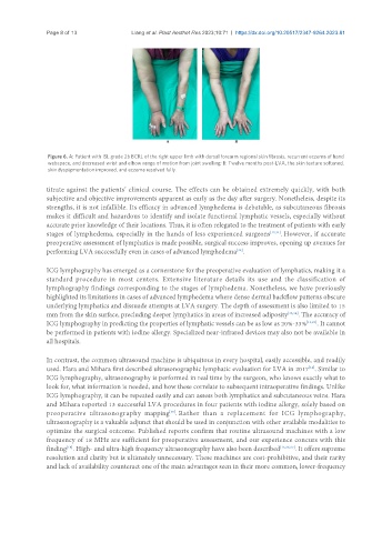
Figure 6. A: Patient with ISL grade 2b BCRL of the right upper limb with dorsal forearm regional skin fibrosis, recurrent eczema of hand
webspace, and decreased wrist and elbow range of motion from joint swelling; B: Twelve months post-LVA, the skin texture softened,
skin dyspigmentation improved, and eczema resolved fully.
titrate against the patients’ clinical course. The effects can be obtained extremely quickly, with both
subjective and objective improvements apparent as early as the day after surgery. Nonetheless, despite its
strengths, it is not infallible. Its efficacy in advanced lymphedema is debatable, as subcutaneous fibrosis
makes it difficult and hazardous to identify and isolate functional lymphatic vessels, especially without
accurate prior knowledge of their locations. Thus, it is often relegated to the treatment of patients with early
stages of lymphedema, especially in the hands of less experienced surgeons [12,31] . However, if accurate
preoperative assessment of lymphatics is made possible, surgical success improves, opening up avenues for
[32]
performing LVA successfully even in cases of advanced lymphedema .
ICG lymphography has emerged as a cornerstone for the preoperative evaluation of lymphatics, making it a
standard procedure in most centers. Extensive literature details its use and the classification of
lymphography findings corresponding to the stages of lymphedema. Nonetheless, we have previously
highlighted its limitations in cases of advanced lymphedema where dense dermal backflow patterns obscure
underlying lymphatics and dissuade attempts at LVA surgery. The depth of assessment is also limited to 15
mm from the skin surface, precluding deeper lymphatics in areas of increased adiposity [33,34] . The accuracy of
ICG lymphography in predicting the properties of lymphatic vessels can be as low as 20%-33% [13,23] . It cannot
be performed in patients with iodine allergy. Specialized near-infrared devices may also not be available in
all hospitals.
In contrast, the common ultrasound machine is ubiquitous in every hospital, easily accessible, and readily
used. Hara and Mihara first described ultrasonographic lymphatic evaluation for LVA in 2017 . Similar to
[14]
ICG lymphography, ultrasonography is performed in real time by the surgeon, who knows exactly what to
look for, what information is needed, and how these correlate to subsequent intraoperative findings. Unlike
ICG lymphography, it can be repeated easily and can assess both lymphatics and subcutaneous veins. Hara
and Mihara reported 13 successful LVA procedures in four patients with iodine allergy, solely based on
preoperative ultrasonography mapping . Rather than a replacement for ICG lymphography,
[35]
ultrasonography is a valuable adjunct that should be used in conjunction with other available modalities to
optimize the surgical outcome. Published reports confirm that routine ultrasound machines with a low
frequency of 18 MHz are sufficient for preoperative assessment, and our experience concurs with this
[16]
finding . High- and ultra-high frequency ultrasonography have also been described [15,36,37] . It offers supreme
resolution and clarity but is ultimately unnecessary. These machines are cost-prohibitive, and their rarity
and lack of availability counteract one of the main advantages seen in their more common, lower-frequency