
Page 11 - Read Online
P. 11
Liang et al. Plast Aesthet Res 2023;10:71 https://dx.doi.org/10.20517/2347-9264.2023.81 Page 5 of 13
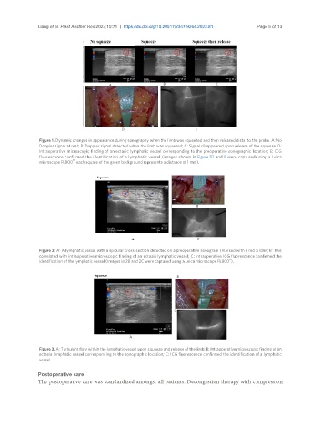
Figure 1. Dynamic changes in appearance during sonography when the limb was squeezed and then released distal to the probe. A: No
Doppler signal at rest; B: Doppler signal detected when the limb was squeezed; C: Signal disappeared upon release of the squeeze; D:
Intraoperative microscopic finding of an ectasic lymphatic vessel corresponding to the preoperative sonographic location; E: ICG
fluorescence confirmed the identification of a lymphatic vessel (images shown in Figure 1D and E were captured using a Leica
®
microscope FL800 ; each square of the green background represents a distance of 1 mm).
Figure 2. A: A lymphatic vessel with a spicular cross-section detected on a preoperative sonogram (marked with a red circle); B: This
correlated with intraoperative microscopic finding of an ectasia lymphatic vessel; C: Intraoperative ICG fluorescence confirmed the
®
identification of the lymphatic vessel (Images in 2B and 2C were captured using a Leica microscope FL800 ).
Figure 3. A: Turbulent flow within the lymphatic vessel upon squeeze and release of the limb; B: Intraoperative microscopic finding of an
ectasia lymphatic vessel corresponding to the sonographic location; C: ICG fluorescence confirmed the identification of a lymphatic
vessel.
Postoperative care
The postoperative care was standardized amongst all patients. Decongestion therapy with compression