
Page 10 - Read Online
P. 10
Grünherz et al. Plast Aesthet Res 2023;10:20 https://dx.doi.org/10.20517/2347-9264.2023.24 Page 5 of 8
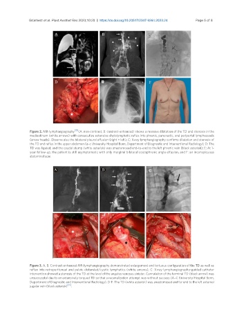
Figure 2. MR-lymphangiography [35] (A: non-contrast; B: contrast-enhanced) shows a massive dilatation of the TD and stenosis in the
mediastinum (white arrows) with consecutive extensive chylolymphatic reflux into phrenic, pancreatic, and periportal lymphvessels
(arrow heads). Observe also the bilateral pleural effusion (right > left); C: X-ray lymphangiography confirms dilatation and stenosis of
the TD and reflux in the upper abdomen (a–c University Hospital Bonn, Department of Diagnostic and Interventional Radiology); D: The
TD was ligated, and the caudal stump (white asterisk) was anastomosed end-to-end to the left phrenic vein (black asterisk); E: At 1-
year follow-up, the patient is still asymptomatic with only marginal bilateral costophrenic angle effusion, and F: an inconspicuous
abdominal scar.
Figure 3. A, B: Contrast-enhanced MR-lymphangiography demonstrated enlargement and tortuous configuration of the TD as well as
reflux into retroperitoneal and pelvic distended/cystic lymphatics (white arrows); C: X-ray lymphangiography-guided catheter
intervention showed a stenosis of the TD at the level of the angulus venosus sinister. Cannulation of the terminal TD (black arrow) was
unsuccessful due to an extensively torqued TD so that a recanalization attempt was without success (A–C University Hospital Bonn,
Department of Diagnostic and Interventional Radiology). D-F: The TD (white asterisk) was anastomosed end-to-end to the left external
jugular vein (black asterisk) [35] .