
Page 62 - Read Online
P. 62
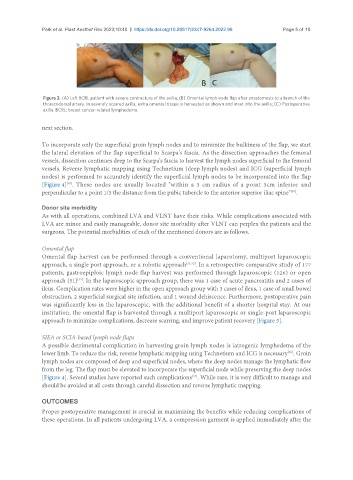
Park et al. Plast Aesthet Res 2023;10:40  https://dx.doi.org/10.20517/2347-9264.2022.98  Page 5 of 10


                Figure 3. (A) Left BCRL patient with severe contracture of the axilla; (B) Omental lymph node flap after anastomosis to a branch of the
                thoracodorsal artery. In severely scarred axilla, extra omental tissue is harvested as shown and inset into the axilla; (C) Postoperative
                axilla. BCRL: breast cancer-related lymphedema.

               next section.


               To incorporate only the superficial groin lymph nodes and to minimize the bulkiness of the flap, we start
               the lateral elevation of the flap superficial to Scarpa's fascia. As the dissection approaches the femoral
               vessels, dissection continues deep to the Scarpa's fascia to harvest the lymph nodes superficial to the femoral
               vessels. Reverse lymphatic mapping using Technetium (deep lymph nodes) and ICG (superficial lymph
               nodes) is performed to accurately identify the superficial lymph nodes to be incorporated into the flap
               [Figure 4] . These nodes are usually located "within a 3 cm radius of a point 3cm inferior and
                       [30]
                                                                                                   [30]
               perpendicular to a point 1/3 the distance from the pubic tubercle to the anterior superior iliac spine" .
               Donor site morbidity
               As with all operations, combined LVA and VLNT have their risks. While complications associated with
               LVA are minor and easily manageable, donor site morbidity after VLNT can perplex the patients and the
               surgeons. The potential morbidities of each of the mentioned donors are as follows.


               Omental flap
               Omental flap harvest can be performed through a conventional laparotomy, multiport laparoscopic
               approach, a single port approach, or a robotic approach [31,32] . In a retrospective comparative study of 177
               patients, gastroepiploic lymph node flap harvest was performed through laparoscopic (126) or open
               approach (51) . In the laparoscopic approach group, there was 1 case of acute pancreatitis and 2 cases of
                           [33]
               ileus. Complication rates were higher in the open approach group with 3 cases of ileus, 1 case of small bowel
               obstruction, 2 superficial surgical site infection, and 1 wound dehiscence. Furthermore, postoperative pain
               was significantly less in the laparoscopic, with the additional benefit of a shorter hospital stay. At our
               institution, the omental flap is harvested through a multiport laparoscopic or single-port laparoscopic
               approach to minimize complications, decrease scarring, and improve patient recovery [Figure 5].

               SIEA or SCIA-based lymph node flaps
               A possible detrimental complication in harvesting groin lymph nodes is iatrogenic lymphedema of the
               lower limb. To reduce the risk, reverse lymphatic mapping using Technetium and ICG is necessary . Groin
                                                                                                  [30]
               lymph nodes are composed of deep and superficial nodes, where the deep nodes manage the lymphatic flow
               from the leg. The flap must be elevated to incorporate the superficial node while preserving the deep nodes
                                                                  [34]
               [Figure 4]. Several studies have reported such complications . While rare, it is very difficult to manage and
               should be avoided at all costs through careful dissection and reverse lymphatic mapping.

               OUTCOMES
               Proper postoperative management is crucial in maximizing the benefits while reducing complications of
               these operations. In all patients undergoing LVA, a compression garment is applied immediately after the