
Page 37 - Read Online
P. 37
Page 4 of 9 Giacalone et al. Plast Aesthet Res 2023;10:22 https://dx.doi.org/10.20517/2347-9264.2022.115
Figure 3. Visualization of lymphatic vessel and vein following ultra-high frequency ultrasound.
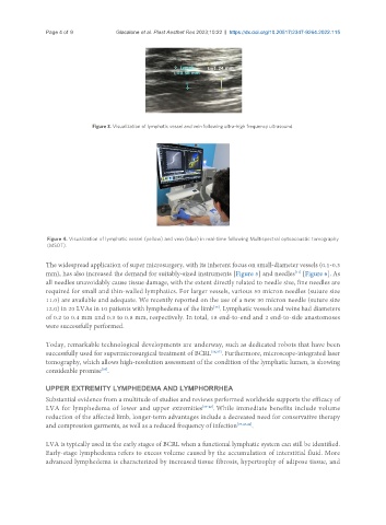
Figure 4. Visualization of lymphatic vessel (yellow) and vein (blue) in real-time following Multispectral optoacoustic tomography
(MSOT).
The widespread application of super microsurgery, with its inherent focus on small-diameter vessels (0.1-0.3
[11]
mm), has also increased the demand for suitably-sized instruments [Figure 5] and needles [Figure 6]. As
all needles unavoidably cause tissue damage, with the extent directly related to needle size, fine needles are
required for small and thin-walled lymphatics. For larger vessels, various 50 micron needles (suture size
11.0) are available and adequate. We recently reported on the use of a new 30 micron needle (suture size
12.0) in 20 LVAs in 10 patients with lymphedema of the limb . Lymphatic vessels and veins had diameters
[35]
of 0.2 to 0.4 mm and 0.3 to 0.8 mm, respectively. In total, 18 end-to-end and 2 end-to-side anastomoses
were successfully performed.
Today, remarkable technological developments are underway, such as dedicated robots that have been
successfully used for supermicrosurgical treatment of BCRL [36,37] . Furthermore, microscope-integrated laser
tomography, which allows high-resolution assessment of the condition of the lymphatic lumen, is showing
consideable promise .
[38]
UPPER EXTREMITY LYMPHEDEMA AND LYMPHORRHEA
Substantial evidence from a multitude of studies and reviews performed worldwide supports the efficacy of
LVA for lymphedema of lower and upper extremities [39-42] . While immediate benefits include volume
reduction of the affected limb, longer-term advantages include a decreased need for conservative therapy
and compression garments, as well as a reduced frequency of infection [39,43,44] .
LVA is typically used in the early stages of BCRL when a functional lymphatic system can still be identified.
Early-stage lymphedema refers to excess volume caused by the accumulation of interstitial fluid. More
advanced lymphedema is characterized by increased tissue fibrosis, hypertrophy of adipose tissue, and