
Page 36 - Read Online
P. 36
Giacalone et al. Plast Aesthet Res 2023;10:22 https://dx.doi.org/10.20517/2347-9264.2022.115 Page 3 of 9
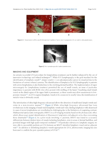
Figure 1. A: Funnelization of the vein; B: Intraluminal insertion of nylon stent in preparation for an end-to-side anastomosis.
Figure 2. Peri-operative proof of patent anastomosis.
IMAGING AND EQUIPMENT
An already successful LVA procedure for lymphedema treatment can be further enhanced by the use of
innovative technology and refined techniques . While ICG lymphography is the gold standard for the
[27]
[28]
identification of lymphatic vessels , deeper vessels (> 2 cm subcutaneously) cannot be visualized due to the
limitations of current infrared cameras. The identification of lymphatics by ICG lymphography in patients
with severe lymphedema is also limited due to the overlying dermal backflow. The implementation of super
microsurgery for lymphedema treatment permitted the use of small vessels, an issue of particular
importance in patients with BCRL who often present with swelling of the hand. Visualizing small lymph
vessels in the distal region of the upper limb is paramount, as these vessels may allow anastomosis to low-
pressure venules . As LVA requires lymphatic vessels to be connected to nearby veins, the identification of
[14]
suitable veins is obviously crucial.
The introduction of ultra-high-frequency ultrasound allows the detection of small-sized lymph vessels and
veins in a non-invasive manner [29-31] [Figure 3]. While ultra-high frequency ultrasound has been
revolutionary in the imaging of small-sized lymphatic vessels and veins, finding lymphatics with ultrasound
in cases of severe lymphedema can nevertheless be challenging due to the limitations of coaptating contrast.
Multispectral optoacoustic tomography (MSOT) is a 3D imaging modality based on the photoacoustic effect
which allows exact spatial identification of (fluorescent) lymphatics and adjacent veins, thus overcoming
[32]
these drawbacks [Figure 4]. In a pilot study involving 11 patients, MSOT was found to accurately
differentiate between distinct types of vessels including lymphatics, even in areas of dermal backflow, and
[33]
provided images with high spatio-temporal resolution . Of particular relevance to lymphatic surgery, we
were able to successfully perform an LVA between an MSOT-identified lymphatic vessel and an adjacent
vein . In addition to identifying lymphatic vessels appropriate for LVA, photoacoustic imaging has also
[33]
been used to confirm the post-operative patency of LVAs .
[34]