
Page 18 - Read Online
P. 18
Page 196 Walker. Neuroimmunol Neuroinflammation 2020;7:194-214 I http://dx.doi.org/10.20517/2347-8659.2020.09
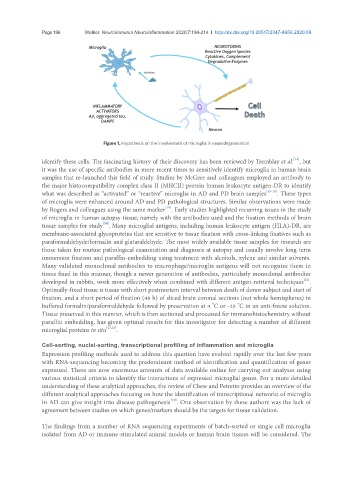
Figure 1. Hypothesis on the involvement of microglia in neurodegeneration
[14]
identify these cells. The fascinating history of their discovery has been reviewed by Tremblay et al. , but
it was the use of specific antibodies in more recent times to sensitively identify microglia in human brain
samples that re-launched this field of study. Studies by McGeer and colleagues employed an antibody to
the major histocompatibility complex class II (MHCII) protein human leukocyte antigen-DR to identify
what was described as “activated” or “reactive” microglia in AD and PD brain samples [15-18] . These types
of microglia were enhanced around AD and PD pathological structures. Similar observations were made
by Rogers and colleagues using the same marker . Early studies highlighted recurring issues in the study
[19]
of microglia in human autopsy tissue, namely with the antibodies used and the fixation methods of brain
tissue samples for study . Many microglial antigens, including human leukocyte antigen (HLA)-DR, are
[20]
membrane-associated glycoproteins that are sensitive to tissue fixation with cross-linking fixatives such as
paraformaldehyde/formalin and glutaraldehyde. The most widely available tissue samples for research are
those taken for routine pathological examination and diagnosis at autopsy and usually involve long-term
immersion fixation and paraffin-embedding using treatment with alcohols, xylene and similar solvents.
Many validated monoclonal antibodies to macrophage/microglia antigens will not recognize them in
tissue fixed in this manner, though a newer generation of antibodies, particularly monoclonal antibodies
[21]
developed in rabbits, work more effectively when combined with different antigen-retrieval techniques .
Optimally-fixed tissue is tissue with short postmortem interval between death of donor subject and start of
fixation, and a short period of fixation (48 h) of sliced brain coronal sections (not whole hemispheres) in
o
o
buffered formalin/paraformaldehyde followed by preservation at 4 C or -20 C in an anti-freeze solution.
Tissue preserved in this manner, which is then sectioned and processed for immunohistochemistry without
paraffin embedding, has given optimal results for this investigator for detecting a number of different
microglial proteins in situ [22,23] .
Cell-sorting, nuclei-sorting, transcriptional profiling of inflammation and microglia
Expression profiling methods used to address this question have evolved rapidly over the last few years
with RNA-sequencing becoming the predominant method of identification and quantification of genes
expressed. There are now enormous amounts of data available online for carrying out analyses using
various statistical criteria to identify the interactions of expressed microglial genes. For a more detailed
understanding of these analytical approaches, the review of Chew and Petretto provides an overview of the
different analytical approaches focusing on how the identification of transcriptional networks of microglia
in AD can give insight into disease pathogenesis . One observation by these authors was the lack of
[10]
agreement between studies on which genes/markers should be the targets for tissue validation.
The findings from a number of RNA sequencing experiments of batch-sorted or single cell microglia
isolated from AD or immune-stimulated animal models or human brain tissues will be considered. The