
Page 10 - Read Online
P. 10
Page 78 Tanaka. Neuroimmunol Neuroinflammation 2020;7:73-91 I http://dx.doi.org/10.20517/2347-8659.2020.04
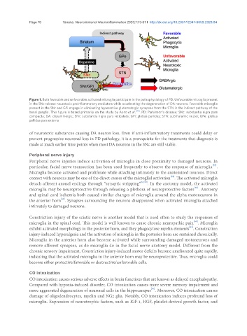
Figure 1. Both favorable and unfavorable activated microglia participate in the pathophysiology of PD. Unfavorable microglia present
in the SNc release neurotoxic proinflammatory mediators while accelerating the degeneration of DA neurons. Favorable microglia
present in the SNr and GPi engage in eliminating hyperactive glutamatergic synapses from the STN in the indirect pathway of the
basal ganglia. This figure is based primarily on the study by Aono et al . [47] . PD: Parkinson’s disease; SNc: substantia nigra pars
compacta; DA: dopaminergic; SNr: substantia nigra pars reticulata; GPi: globus pallidus; STN: subthalamic nuclei; GPe: globus
pallidus pars externa
of neurotoxic substances causing DA neuron loss. Even if anti-inflammatory treatments could delay or
prevent progressive neuronal loss in PD pathology, it is a prerequisite for the treatments that diagnosis is
made at much earlier time points when most DA neurons in the SNc are still viable.
Peripheral nerve injury
Peripheral nerve injuries induce activation of microglia in close proximity to damaged neurons. In
[57]
particular, facial nerve transection has been used frequently to observe the response of microglia .
Microglia become activated and proliferate while attaching intimately to the axotomized neurons. Direct
[58]
contact with neurons may be one of the direct causes of the microglial activation . The activated microglia
detach afferent axonal endings through “synaptic stripping” [57,59] . In the axotomy model, the activated
[60]
microglia may be neuroprotective through releasing a plethora of neuroprotective factors . Axotomy
and spinal cord ischemia both caused similar changes of microglia around the alpha motoneurons in
[61]
the anterior horn . Synapses surrounding the neurons disappeared when activated microglia attached
intimately to damaged neurons.
Constriction injury of the sciatic nerve is another model that is used often to study the responses of
[62]
microglia in the spinal cord. This model is well known to cause chronic neuropathic pain . Microglia
[63]
exhibit activated morphology in the posterior horn, and they phagocytose myelin elements . Constriction
injury-induced hyperalgesia and the activation of microglia in the posterior horn are sustained chronically.
Microglia in the anterior horn also become activated while surrounding damaged motoneurons and
remove afferent synapses, as do microglia do in the facial nerve axotomy model. Different from the
chronic sensory impairment, Constriction injury-induced motor deficits become ameliorated quite rapidly,
indicating that the activated microglia in the anterior horn may be neuroprotective. Thus, microglia could
become either protective/favorable or destructive/unfavorable cells.
CO intoxication
CO intoxication causes serious adverse effects in brain functions that are known as delayed encephalopathy.
Compared with hypoxia-induced disorder, CO intoxication causes more severe memory impairment and
more aggravated degeneration of neuronal cells in the hippocampus . Moreover, CO intoxication causes
[64]
damage of oligodendrocytes, myelin and NG2 glia. Notably, CO intoxication induces profound loss of
microglia. Expression of neurotrophic factors, such as IGF-1, HGF, platelet-derived growth factor, and