Page 167 - Read Online
P. 167
Page 6 of 24                                 Neuroimmunol Neuroinflammation 2019;6:15  I  http://dx.doi.org/10.20517/2347-8659.2019.019
                                           -
                              3+
                 2+
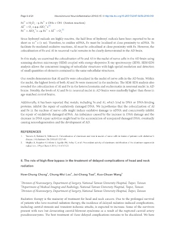
               Fe  + H O  → Fe  + OH• + OH  (Fenton reaction)
                        2
                      2
               Al  + O • ↔ AlO •
                 3+
                                2+ [2]
                       -
                      2
                               2
                                2+
                         2+
                 3+
                                      3+
               Fe  + AlO • → Fe  + Al  + O 2 [2]
                        2
               Since hydroxyl radicals are highly reactive, the half-lives of hydroxyl radicals have been reported to be as
                         -9
               short as 10  s (1 ns). Therefore, to oxidize nDNA, Fe must be localized at close proximity to nDNA. To
               facilitate Fe-mediated oxidative reactions, Al must be colocalized at close proximity with Fe. However, the
               colocalization of Fe and Al in neuronal nuclei remains to be clearly demonstrated in the AD brain.
               In this study, we examined the colocalization of Fe and Al in the nuclei of nerve cells in the AD brain using
               scanning electron microscopy (SEM) coupled with energy-dispersive X-ray spectroscopy (EDS). SEM-EDS
               analysis allows the concurrent imaging of subcellular structures with high spatial resolution and detection
               of small quantities of elements contained in the same subcellular structures.
               Our results demonstrate that Al and Fe were colocalized in the nuclei of nerve cells in the AD brain. Within
               the nuclei, the highest levels of both Al and Fe were measured in the nucleolus. The SEM-EDS analysis also
               revealed the colocalization of Al and Fe in the heterochromatin and euchromatin in neuronal nuclei in AD
               brains. Notably, the levels of Al and Fe in neuronal nuclei in AD brains were markedly higher than those in
               age-matched control brains.

               Additionally, it has been reported that metals, including Fe and Al, which bind to DNA or DNA-binding
               proteins, inhibit the repair of oxidatively damaged DNA. We hypothesize that the colocalization of Al
               and Fe in the nucleus of nerve cells might induce oxidative damage to nDNA and concurrently inhibit
               the repair of oxidatively damaged nDNA. An imbalance caused by the increase in DNA damage and the
               decrease in DNA repair activities might lead to the accumulation of unrepaired damaged DNA, eventually
               causing neurodegeneration and the development of AD.


               REFERENCES
               1.   Yumoto S, Kakimi S, Ishikawa A. Colocalization of aluminum and iron in nuclei of nerve cells in brains of patients with alzheimer’s
                   disease. J Alzheimers Dis 2018;65:1267-81.
               2.   Mujika JI, Ruipérez F, Infante I, Ugalde JM, Exley C, et al. Pro-oxidant activity of aluminum: stabilization of the aluminum superoxide
                   radical ion. J Phys Chem A 2011;115:6717-23.


               8. The role of high-flow bypass in the treatment of delayed complications of head and neck

               radiation

                                 1
                                                 2
                                                                 3
               How-Chung Cheng , Chung-Wei Lee , Jui-Chang Tsai , Kuo-Chuan Wang    3
               1 Division of Neurosurgery, Department of Surgery, National Taiwan University Hospital, Taipei, Taiwan
               2 Department of Medical Imaging and Radiology, National Taiwan University Hospital, Taipei, Taiwan
               3 Division of Neurosurgery, Department of Surgery, National Taiwan University Hospital, Taipei, Taiwan

               Radiation therapy is the mainstay of treatment for head and neck cancers. Due to the prolonged survival
               of patients who have received radiation therapy, the incidence of delayed radiation-induced complications,
               including carotid stenosis and transient ischemic attacks, is expected to increase. Some of the survivors
               present with rare but devastating carotid blowout syndrome as a result of the ruptured carotid artery
               pseudoaneurysms. The best treatment of these delayed complications remains to be elucidated. We here

