
Page 195 - Read Online
P. 195
Dai et al. Neuroimmunol Neuroinflammation 2018;5:28 I http://dx.doi.org/10.20517/2347-8659.2018.09 Page 3 of 8
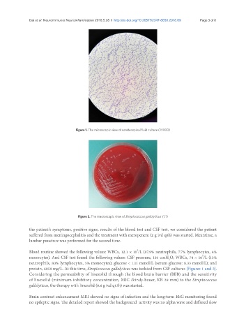
Figure 1. The microscopic view of cerebrospinal fluid culture (1:1000)
Figure 2. The macroscopic view of Streptococcus gallolyticus (1:1)
the patient’s symptoms, positive signs, results of the blood test and CSF test, we considered the patient
suffered from meningocephalitis and the treatment with meropenem (2 g ivd q8h) was started. Meantime, a
lumbar puncture was performed for the second time.
9
Blood routine showed the following values: WBCs, 12.1 × 10 /L (87.9% neutrophils, 7.7% lymphocytes, 4%
6
monocytes). And CSF test found the following values: CSF pressure, 110 cmH O; WBCs, 74 × 10 /L (15%
2
neutrophils, 80% lymphocytes, 5% monocytes); glucose < 1.11 mmol/L (serum glucose: 8.33 mmol/L); and
protein, 6058 mg/L. At this time, Streptococcus gallolyticus was isolated from CSF cultures [Figures 1 and 2].
Considering the permeability of linezolid through the blood brain barrier (BBB) and the sensitivity
of linezolid (minimum inhibitory concentration, MIC /kindy-bauer, KB 29 mm) to the Streptococcus
gallolyticus, the therapy with linezolid (0.6 g ivd q12h) was started.
Brain contrast enhancement MRI showed no signs of infection and the long-term EEG monitoring found
no epileptic signs. The detailed report showed the background activity was no alpha wave and diffused slow