
Page 52 - Read Online
P. 52
Page 6 of 11 Kato et al. Mini-invasive Surg 2021;5:5 I http://dx.doi.org/10.20517/2574-1225.2020.98
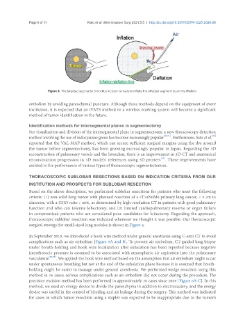
Figure 3. The targeted segmental bronchus incision is made to inflate the affected segment by air insufflation.
embolism by avoiding parenchymal puncture. Although these methods depend on the equipment of every
institution, it is expected that an iVATS method or a wireless marking system will become a significant
method of tumor identification in the future.
Identification methods for intersegmental planes in segmentectomy
For visualization and division of the intersegmental plane in segmentectomy, a new thoracoscopy detection
method involving the use of indocyanine green has become increasingly popular [49-51] . Furthermore, Sato et al. [52]
reported that the VAL-MAP method, which can secure sufficient surgical margins using the dye around
the tumor before segmentectomy, has been growing increasingly popular in Japan. Regarding the 3D
reconstruction of pulmonary vessels and the bronchus, there is an improvement in 3D-CT and anatomical
[53]
reconstruction progression in 3D models’ references using 3D printers . These improvements have
assisted in the performance of various types of thoracoscopic segmentectomies.
THORACOSCOPIC SUBLOBAR RESECTIONS BASED ON INDICATION CRITERIA FROM OUR
INSTITUTION AND PROSPECTS FOR SUBLOBAR RESECTION
Based on the above description, we performed sublobar resections for patients who meet the following
criteria: (1) non-solid lung tumor with planned resection of a cT1aN0M0 primary lung cancer, < 2 cm in
diameter, with a GGO ratio > 80%, as determined by high-resolution CT in patients with good pulmonary
function and who can tolerate lobectomy; and (2) limited cardiopulmonary reserve or organ failure
in compromised patients who are considered poor candidates for lobectomy. Regarding the approach,
thoracoscopic sublobar resection was indicated whenever we thought it was possible. Our thoracoscopic
surgical strategy for small-sized lung nodules is shown in Figure 4.
In September 2015, we introduced a hook wire method under general anesthesia using C-arm CT to avoid
complications such as air embolism [Figure 5A and B]. To prevent air embolism, CT-guided lung biopsy
under breath-holding and hook wire localization after exhalation has been reported because negative
intrathoracic pressure is assumed to be associated with atmospheric air aspiration into the pulmonary
vasculature [54-56] . We applied the hook wire method based on the assumption that air embolism might occur
under spontaneous breathing but not at the end of the exhalation phase because it is assumed that breath-
holding might be easier to manage under general anesthesia. We performed wedge resection using this
method in 16 cases; serious complications such as air embolism did not occur during the procedure. The
precision excision method has been performed in approximately 20 cases since 2009 [Figure 6A-C]. In this
method, we used an energy device to divide the parenchyma in addition to electrocautery, and the energy
device was useful in the control of bleeding and air leakage during the surgery. This method was indicated
for cases in which tumor resection using a stapler was expected to be inappropriate due to the tumor’s