
Page 51 - Read Online
P. 51
Kato et al. Mini-invasive Surg 2021;5:5 I http://dx.doi.org/10.20517/2574-1225.2020.98 Page 5 of 11
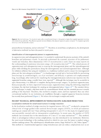
Figure 2. Slip-knot technique. The slip-knot made with a monofilament thread is fully pulled to ligate the targeted segmental bronchus
after bilateral lung ventilation. The affected segment remains inflated, while adjacent segments appear collapsed. The targeted segmental
bronchus is then divided with a stapler.
pneumothorax, hematoma, and air embolism [37,38] . Therefore, to avoid these complications, the development
of alternative methods has been discussed in recent years.
Identification of intersegmental planes in segmentectomy
In segmentectomy and subsegmentectomy, it is essential to understand the precise anatomy of the patient’s
bronchus and pulmonary vessels. To precisely understand the anatomic structure of the pulmonary
vessels and bronchus, three-dimensional (3D) CT reconstruction is used. There are many reports on
the understanding of anatomical structures using a 3D reconstruction tool [39-41] . The critical process of
segmentectomy and subsegmentectomy is ensuring the intersegmental plane and the intersubsegmental
plane while dividing the parenchyma along the intersegmental and intersubsegmental lines. Although
the inflation-deflation line using jet ventilation is a traditional method used to ensure the intersegmental
[42]
plane and the intersubsegmental planes , its disadvantages include lack of technical skills for performing
bronchoscopy by anesthesiologists, use of jet ventilation, and difficulty in patients with emphysematous
lung. Accordingly, other methods for visualizing the intersegmental plane (selective dye injection into the
[43]
segmental bronchus using a needle) have been reported . Although this technique may be accessible in
open thoracotomy, it is difficult to complete during thoracoscopic surgery. To perform segmentectomy
thoracoscopically, we have improved thoracoscopic segmentectomy using the following simplified
[44]
technique: the slip-knot technique for creating an intersegmental plane [Figure 2] . The essential device
of this technique is simply a slip-knot made by a monofilament thread, and the essential process is merely
pulling of the slip-knot. Therefore, this method is simpler, easier, and less expensive than any other
conventional method. In our institution, air insufflation through a targeted segmental bronchus incision
has recently been performed [Figure 3]. We believe this technique is simple and useful.
RECENT TECHNICAL IMPROVEMENTS IN THORACOSCOPIC SUBLOBAR RESECTIONS
Localization methods for small-sized tumors in wedge resection
[45]
In the localization method of wedge resection for targeted tumors, Gill et al. conducted a prospective
clinical trial of image-guided video-assisted thoracoscopic surgery (iVATS), in which percutaneous
markings are created with two T-bars utilizing intraoperative C-arm CT. In this study, the targeted tumor
was successfully resected with no intraoperative complications. In recent years, the number of iVATS
methods has increased due to the introduction of C-arm CT in many institutions [46,47] . This method,
which is advantageous without serious complications, was also introduced to our institution. Moreover,
the most recent technology is the marking method in which the area near the tumor is marked using a
[48]
wireless marking system (The radiofrequency identification system, Hogy Medical Co, Ltd, Tokyo, Japan) .
This method is a transbronchial approach using bronchoscopy and can reduce complications such as air