
Page 438 - Read Online
P. 438
Page 4 of 14 Choi et al. Mini-invasive Surg 2021;5:43 https://dx.doi.org/10.20517/2574-1225.2021.73
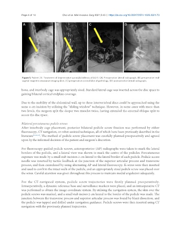
Figure 1. Patient 24. Treatment of degenerative spondylolisthesis of L4/5. (A) Preoperative lateral radiograph, (B) preoperative mid-
sagittal magnetic resonance imaging slice, (C) preoperative coronal slice at pathology, (D) postoperative lateral radiograph.
bone, and interbody cage was appropriately sized. Standard lateral cage was inserted across the disc space to
gaining bilateral cortical endplate coverage.
Due to the mobility of the abdominal wall, up to three intervertebral discs could be approached using the
same 4 cm incision by utilising the “sliding window” technique. However, in some cases with more than
two levels, the surgeon split the deeper two muscles twice, having extended the external oblique split to
access the disc space.
Bilateral percutaneous pedicle screws
After interbody cage placement, posterior bilateral pedicle screw fixation was performed by either
fluoroscopy, CT navigation, or robot-assisted techniques, all of which have been previously described in the
literature [17,30,31] . The method of pedicle screw placement was carefully planned preoperatively and agreed
upon by the informed decision of the patient and surgeon’s discretion.
For fluoroscopy-guided pedicle screws, anteroposterior (AP) radiographs were taken to mark the lateral
borders of the pedicle, and a lateral view was shown to mark the centre of the pedicles. Percutaneous
exposure was made by a small stab incision 2 cm lateral to the lateral border of each pedicle. Pedicle access
needle was inserted by tactile feedback at the junction of the superior articular process and transverse
process, and then cannulated by using alternating AP and lateral fluoroscopy. K-wires were then inserted
and used to confirm the intact walls of the pedicle, and an appropriately sized pedicle screw was placed over
the wires. Careful attention was given throughout this process to maintain medial angulation adequately.
For the CT-navigated system, pedicle screw trajectories were firstly planned preoperatively.
Intraoperatively, a dynamic reference base and surveillance markers were placed, and an intraoperative CT
was performed to obtain the image coordinate system. By utilising the navigation system, the skin over the
pedicle screws was marked, and a small stab incises 2 cm lateral to the border of the pedicle was made. The
junction between the transverse process and superior articular process was found by blunt dissection, and
the pedicle was tapped and drilled under navigation guidance. Pedicle screws were then inserted using CT
navigation with the previously planned trajectories.