
Page 358 - Read Online
P. 358
Bianchi et al. Mini-invasive Surg 2021;5:37 https://dx.doi.org/10.20517/2574-1225.2021.64 Page 3 of 11
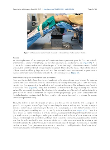
Figure 1. Full flank position and bent bed to increase the distance between the iliac crest and the ribs.
Access
To identify placement of the camera port and creation of the retroperitoneal space, the iliac crest, 12th rib,
and the inferior lumbar (Petit’s) triangle are important landmarks and can be marked out [Figure 2A]. A 1.5
cm vertical incision is made at the level of the apex of the Petit’s triangle. Subcutaneous tissue is divided
with cautery until the internal oblique muscle is reached. Thereafter, the muscle fibers of the internal
oblique muscle are bluntly finger-separated, and then Metzenbaum scissors are used to penetrate the
thoracolumbar and trasversalis fasciae and enter the retroperitoneal space [Figure 2B].
Retroperitoneal space creation and port placement
After inserting the index finger into the previous incision, the retroperitoneal space between the posterior
layer of renal fascia and the transversalis fascia is created by turning the index finger in a 180° movement,
running it as close as possible to the abdominal wall, separating the pararenal fat and peritoneum from the
transversalis fascia [Figure 3]. During this maneuver, the sensation of the finger running on a smooth
surface (the transversalis fascia) and the palpation of the internal surface of the 12th rib and the body of the
psoas muscle are crucial to ensure that the surgeon is developing the right space. In case these internal and
haptic landmarks are not perceived, the finger could be in the wrong place, such as in between the muscles
or inside the peritoneal cavity.
Then, the first two 8 mm robotic ports are placed at a distance of 8 cm from the first access port - it
generally corresponds to one-finger length - one along the anterior axillary line, the other along the
posterior axillary line, 1-2 cm cranially to the level of the camera port. A 12 mm AirSeal® assistant port is
placed on the posterior axillary line, 3-5 cm caudally to the 8 mm robotic port [Figure 2C-E]. These first
three trocars are bluntly positioned in a “blind fashion”, keeping the index finger through the first access
port inside the retroperitoneal space, pushing on the abdominal wall at the site of trocar insertions. In this
way, the positioning is both fast and safe, although blind, because the internal finger guarantees that nothing
else than the abdominal wall is along the route of the trocar. After the insertion of the two first 8 mm
robotic trocars and the AirSeal® trocar, the 8 mm robotic camera port, through a Hasson cone, is placed in
the first incision. Then, pneumoretroperitoneum is created at 12 mmHg of carbon dioxide and the 0°
robotic camera can be inserted in the retroperitoneal cavity.