
Page 630 - Read Online
P. 630
Dharmaraj et al. Mini-invasive Surg 2020;4:65 I http://dx.doi.org/10.20517/2574-1225.2020.51 Page 3 of 6
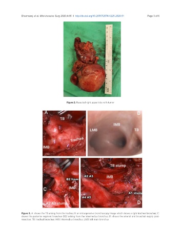
Figure 2. Resected right upper lobe with tumor
Figure 3. A: shows the TB arising from the trachea; B: an intraoperative bronchoscopy image which shows a right tracheal bronchus; C:
shows the posterior segment bronchus (B2) arising from the intermedius bronchus; D: shows the arterial and bronchial supply post-
resection. TB: tracheal bronchus; IMB: intermedius bronchus; LMB: left main bronchus