
Page 197 - Read Online
P. 197
Page 4 of 11 Cicero et al. Mini-invasive Surg 2019;3:25 I http://dx.doi.org/10.20517/2574-1225.2018.012
A B
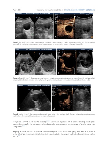
Figure 3. Bosniak 2F cyst. A: Grey scale sonography shows in the mid portion of the left kidney cystic lesion with thin hyperechoic
septum; B: Contrast enhanced sonography reveal homogeneous enhancement of the septum without parietal nodule
A B
Figure 4. Bosniak 3 cyst. A: Greyscale sonography shows voluminous mass with mixed echo structure (anechoic and hyperechoic
components); B: Contrast enhanced sonography reveals cystic lesion characterized by irregular, thickened and vascularized walls
A B
Figure 5. Bosniak 4 cyst. A: Grey scale shows hypoechoic round lesion with smooth margin; B: Contrast- enhanced sonography reveals a
cystic lesion with mural nodules characterized by strong enhancement
recognize LE with inconclusive findings [11-13] . CEUS has a greater SP in characterizing renal cystic
lesions, in particular the presence and thickness of a septum and/or the presence of a solid intracystic
components [14-16] .
Anyway, it is well-known the role of CT in the malignant cystic lesion for staging: now the CEUS is useful
in the follow up of complex cystic lesions that are not suitable for surgery and in the future it could replace
[15]
the CT .