
Page 165 - Read Online
P. 165
Parthasarathi et al. Mini-invasive Surg 2019;3:20 I http://dx.doi.org/10.20517/2574-1225.2019.10 Page 9 of 14
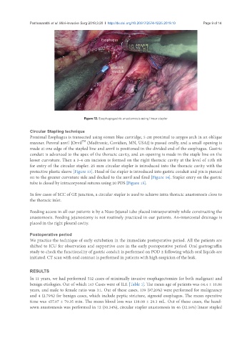
Figure 12. Esophagogastric anastomosis using linear stapler
Circular Stapling technique
Proximal Esophagus is transected using 60mm blue cartridge, 5 cm proximal to azygos arch in an oblique
TM
manner. Peroral anvil [Orvil (Medtronic, Covidien, MN, USA)] is passed orally, and a small opening is
made at one edge of the stapled line and anvil is positioned in the divided end of the esophagus. Gastric
conduit is advanced to the apex of the thoracic cavity, and an opening is made in the staple line on the
lesser curvature. Then a 3-4 cm incision is formed on the right thoracic cavity at the level of 11th rib
for entry of the circular stapler. 25 mm circular stapler is introduced into the thoracic cavity with the
protective plastic sleeve [Figure 13]. Head of the stapler is introduced into gastric conduit and pin is pierced
on to the greater curvature side and docked to the anvil and fired [Figure 14]. Stapler entry on the gastric
tube is closed by intracorporeal sutures using 20 PDS [Figure 15].
In few cases of SCC of GE junction, a circular stapler is used to achieve intra thoracic anastomosis close to
the thoracic inlet.
Feeding access in all our patients is by a Naso-Jejunal tube placed intraoperatively while constructing the
anastomosis. Feeding jejunostomy is not routinely practiced in our patients. An-intercostal drainage is
placed in the right pleural cavity.
Postoperative period
We practice the technique of early extubation in the immediate postoperative period. All the patients are
shifted to ICU for observation and supportive care in the early postoperative period. Oral gastrograffin
study to check the functionality of gastric conduit is performed on POD 2 following which oral liquids are
initiated. CT scan with oral contrast is performed in patients with high suspicion of the leak.
RESULTS
In 11 years, we had performed 532 cases of minimally invasive esophagectomies for both malignant and
benign etiologies. Out of which 143 Cases were of ILE [Table 1]. The mean age of patients was 64.4 ± 10.86
years, and male to female ratio was 3:1. Out of these cases, 139 (97.20%) were performed for malignancy
and 4 (2.79%) for benign cases, which include peptic stricture, sigmoid esophagus. The mean operative
time was 457.97 ± 79.35 min. The mean blood loss was 138.08 ± 29.3 mL. Out of these cases, the hand-
sewn anastomosis was performed in 72 (50.34%), circular stapler anastomosis in 46 (32.16%) linear stapled