
Page 105 - Read Online
P. 105
Mazzola et al. Mini-invasive Surg 2019;3:12 I http://dx.doi.org/10.20517/2574-1225.2019.05 Page 3 of 10
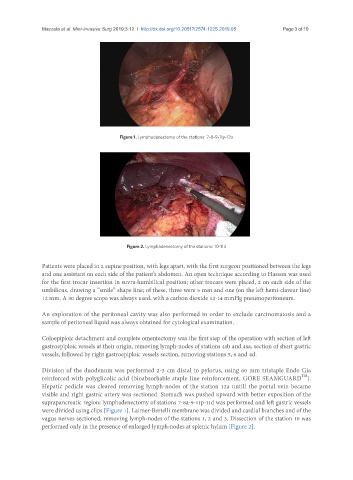
Figure 1. Lymphadenectomy of the stations: 7-8-9-11p-12a
Figure 2. Lymphadenectomy of the stations: 10-11d
Patients were placed in a supine position, with legs apart, with the first surgeon positioned between the legs
and one assistant on each side of the patient’s abdomen. An open technique according to Hasson was used
for the first trocar insertion in suvra-humbilical position; other trocars were placed, 2 on each side of the
umbilicus, drawing a “smile” shape line; of these, three were 5 mm and one (on the left hemi-clavear line)
12 mm. A 30 degree scope was always used, with a carbon dioxide 12-14 mmHg pneumoperitoneum.
An exploration of the peritoneal cavity was also performed in order to exclude carcinomatosis and a
sample of peritoneal liquid was always obtained for cytological examination.
Coloepiploic detachment and complete omentectomy was the first step of the operation with section of left
gastroepiploic vessels at their origin, removing lymph-nodes of stations 4sb and 4sa, section of short gastric
vessels, followed by right gastroepiploic vessels section, removing stations 5, 6 and 4d.
Division of the duodenum was performed 2-3 cm distal to pylorus, using 60 mm tristaple Endo Gia
TM
reinforced with polyglicolic acid (bioabsorbable staple line reinforcement, GORE SEAMGUARD ).
Hepatic pedicle was cleared removing lymph-nodes of the station 12a untill the portal vein became
visible and right gastric artery was sectioned. Stomach was pushed upward with better exposition of the
suprapancreatic region; lymphadenectomy of stations 7-8a-9-11p-11d was performed and left gastric vessels
were divided using clips [Figure 1]. Laimer-Bertelli membrane was divided and cardial branches and of the
vagus nerves sectioned, removing lymph-nodes of the stations 1, 2 and 3. Dissection of the station 10 was
performed only in the presence of enlarged lymph-nodes at splenic hylum [Figure 2].