
Page 285 - Read Online
P. 285
Page 4 of 6 Ke et al. Mini-invasive Surg 2018;2:35 I http://dx.doi.org/10.20517/2574-1225.2018.46
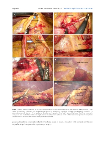
Figure 2. Splenic flexure mobilization. A: Entering the lesser sac by dividing the mesentery at the inferior border of the pancreas; B: high
ligation of the inferior mesenteric vein proximal to the ligament of Treitz and on the inferior border of the pancreas; C: division of the
pancreaticomesocolic ligament; D: retroperitoneal dissection and separation of the Toldt’s fascia; E: ligation of the inferior mesenteric
artery at its root; F: lateral dissection involving division of the left paracolic gutter; G: division of the splenocolic ligament; H: completion
of splenic flexure mobilization by division of the gastrocolic ligaments
proach utilized is a combined medial to lateral and lateral to medial dissection with emphasis on the ease
of performing the steps during laparoscopic surgery.