
Page 11 - Read Online
P. 11
Palma et al. Mini-invasive Surg 2018;2:1 I http://dx.doi.org/10.20517/2574-1225.2017.40 Page 3 of 5
Figure 2. The nematode removal by biopsy forceps
Figure 3. Transverse section of Anisakis type I larva in the gastric mucosa. Scale bar 100 mm
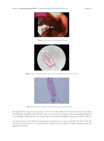
Figure 4. Sagittal section of Anisakis type I larva in the gastric mucosa. Magnification 40x
the sagittal section [Figure 4] two glandular cells found in the caudal end of the nematode were also visible.
No ventricular appendix and/or intestinal caecum were found. According to those morphological features
it was possible to refer the worm to a larval stage of a nematode belonging to the genus Anisakis [Video 1].
The patient reported she had eaten marinated raw anchovies at a sushi restaurant five days before the
procedure and she described an intensification of gastric pain, in absence of other symptoms, after the
ingestion of raw fish.