
Page 10 - Read Online
P. 10
Page 2 of 5 Palma et al. Mini-invasive Surg 2018;2:1 I http://dx.doi.org/10.20517/2574-1225.2017.40
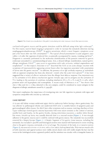
Figure 1. The Anisakis larva visualization in the gastric fundus during retrovision maneuver close to the mucosal erosion
[1]
confused with gastric mucus and the gastric detection could be difficult using white light endoscopy .
For this reason, narrow band imaging is proposed in order to increase the nematode detection during
[2]
esophagogastroduodenoscopy (EGD) . In gastric anisakiasis, which is more frequent, symptoms occur
[3,4]
just a few hours after raw fish consumption . The typical clinical presentation is acute epigastric pain
[5]
within 12 h after the ingestion of infected fish, occasionally accompanied by nausea, vomiting and fever .
Diagnosis is currently performed by the molecular identification of the parasite removed by gastric
endoscopy associated to a seroimmunological assay. Also, a delayed allergic manifestation, named gastro-
[6,7]
allergic anisakiasis (GAA) , may occur in association with rash, urticaria, isolated angioedema and
[8]
[6]
anaphylaxis . In this respect, Daschner et al. described the GAA as an acute allergic reaction with
symptoms of hypersensitivity appearing several hours after the ingestion associated with penetration
[9]
of larvae into the gastric mucosa . In addition, the presence of an IgE antibody response in individuals
[10]
with no apparent symptoms has been also detected 1 month after the acute GAA episode . It has been
suggested that a variety of effector molecules form the allergic host defence response. One important step
in the activation of this defence mechanism against Anisakis spp. and its antigens are the activation of
Th2, leading to the secretion of cytokines, including interleukin (IL)-4, IL-5, IL-9 and IL-13 cells, which
[11]
promote mast cell hyperplasia and eosinophilia. A recent study of Mattiucci et al. has shown that
Ani s 7-like and Ani s 13-like, detected in the sera samples, could be considered as major antigens in the
diagnosis of allergic anisakiasis caused by A. pegreffii.
This report emphasizes the importance of investigating into raw fish ingestion in patients with signs and
symptoms compatible with Anisakis sp. infection.
CASE REPORT
A 52-year-old Italian woman underwent upper elective endoscopy before having a sleeve gastrectomy. She
was affected by pathological obesity and underwent EGD after 12 months history of epigastric pain and
gastroesophageal reflux disease. She didn’t have other symptoms such as nausea, vomiting, diarrhea or fever.
Upper endoscopy showed gastric mucosa hyperemia with some micro-erosions in the context of antrum,
middle body and fundus. In the gastric fundus, during retrovision maneuver, a mobile white thread-
like worm, Anisakis sp. larva, was casually detected close to a mucosal erosion [Figure 1]. It was strongly
adherent to the gastric mucosa and it could be confused with gastric mucus. The nematode was successfully
removed by a biopsy forceps [Figure 2]. A bioptic fragment of the gastric tissue mucosa, including the
nematode, was included in paraffin and histological sections were stained with haematoxylin-eosin revealing
close to the gastric mucosa a nematode, at its larval stage. The diameter of the body, in a transerve section
[Figure 3], was around 0.30 mm × 0.20 mm, with a thin cuticle, lacking lateral alae. Polymiarian muscle cells
at both transverse and sagittal sections were visible; lateral chords were still visible despite the fact that the
worm in the histological section appeared spoiled, and the intestine was circular with a triangular lumen. In