
Page 55 - Read Online
P. 55
Page 4 of 18 Ninomiya et al. Mini-invasive Surg 2022;6:33 https://dx.doi.org/10.20517/2574-1225.2022.12
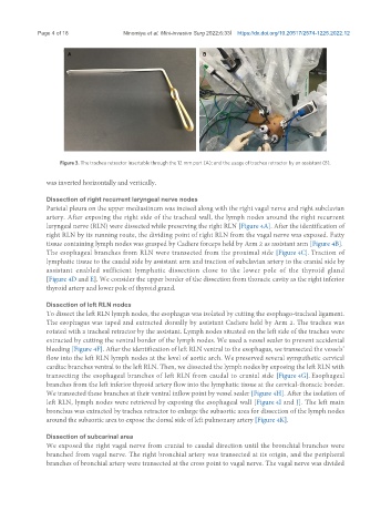
Figure 3. The trachea retractor insertable through the 12 mm port (A); and the usage of trachea retractor by an assistant (B).
was inverted horizontally and vertically.
Dissection of right recurrent laryngeal nerve nodes
Parietal pleura on the upper mediastinum was incised along with the right vagal nerve and right subclavian
artery. After exposing the right side of the tracheal wall, the lymph nodes around the right recurrent
laryngeal nerve (RLN) were dissected while preserving the right RLN [Figure 4A]. After the identification of
right RLN by its running route, the dividing point of right RLN from the vagal nerve was exposed. Fatty
tissue containing lymph nodes was grasped by Cadiere forceps held by Arm 2 as assistant arm [Figure 4B].
The esophageal branches from RLN were transected from the proximal side [Figure 4C]. Traction of
lymphatic tissue to the caudal side by assistant arm and traction of subclavian artery to the cranial side by
assistant enabled sufficient lymphatic dissection close to the lower pole of the thyroid gland
[Figure 4D and E]. We consider the upper border of the dissection from thoracic cavity as the right inferior
thyroid artery and lower pole of thyroid grand.
Dissection of left RLN nodes
To dissect the left RLN lymph nodes, the esophagus was isolated by cutting the esophago-tracheal ligament.
The esophagus was taped and extracted dorsally by assistant Cadiere held by Arm 2. The trachea was
rotated with a tracheal retractor by the assistant. Lymph nodes situated on the left side of the trachea were
extracted by cutting the ventral border of the lymph nodes. We used a vessel sealer to prevent accidental
bleeding [Figure 4F]. After the identification of left RLN ventral to the esophagus, we transected the vessels’
flow into the left RLN lymph nodes at the level of aortic arch. We preserved several sympathetic cervical
cardiac branches ventral to the left RLN. Then, we dissected the lymph nodes by exposing the left RLN with
transecting the esophageal branches of left RLN from caudal to cranial side [Figure 4G]. Esophageal
branches from the left inferior thyroid artery flow into the lymphatic tissue at the cervical-thoracic border.
We transected these branches at their ventral inflow point by vessel sealer [Figure 4H]. After the isolation of
left RLN, lymph nodes were retrieved by exposing the esophageal wall [Figure 4I and J]. The left main
bronchus was extracted by trachea retractor to enlarge the subaortic area for dissection of the lymph nodes
around the subaortic area to expose the dorsal side of left pulmonary artery [Figure 4K].
Dissection of subcarinal area
We exposed the right vagal nerve from cranial to caudal direction until the bronchial branches were
branched from vagal nerve. The right bronchial artery was transected at its origin, and the peripheral
branches of bronchial artery were transected at the cross point to vagal nerve. The vagal nerve was divided