
Page 55 - Read Online
P. 55
Page 4 of 19 Masiero et al. Mini-invasive Surg 2022;6:4 https://dx.doi.org/10.20517/2574-1225.2021.104
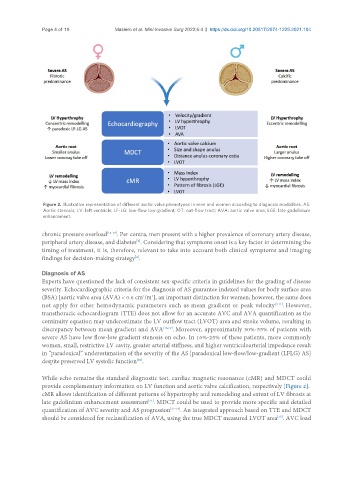
Figure 2. Illustrative representation of different aortic valve phenotypes in men and women according to diagnosis modalities. AS:
Aortic stenosis; LV: left ventricle; LF-LG: low-flow low-gradient; OT: out-flow tract; AVA: aortic valve area; LGE: late gadolinium
enhancement.
chronic pressure overload [15-17] . Per contra, men present with a higher prevalence of coronary artery disease,
[8]
peripheral artery disease, and diabetes . Considering that symptoms onset is a key factor in determining the
timing of treatment, it is, therefore, relevant to take into account both clinical symptoms and imaging
findings for decision-making strategy .
[4]
Diagnosis of AS
Experts have questioned the lack of consistent sex-specific criteria in guidelines for the grading of disease
severity. Echocardiographic criteria for the diagnosis of AS guarantee indexed values for body surface area
2
2
(BSA) [aortic valve area (AVA) < 0.6 cm /m ], an important distinction for women; however, the same does
not apply for other hemodynamic parameters such as mean gradient or peak velocity [4,11] . However,
transthoracic echocardiogram (TTE) does not allow for an accurate AVC and AVA quantification as the
continuity equation may underestimate the LV outflow tract (LVOT) area and stroke volume, resulting in
discrepancy between mean gradient and AVA [18,19] . Moreover, approximately 30%-55% of patients with
severe AS have low flow-low gradient stenosis on echo. In 10%-25% of these patients, more commonly
women, small, restrictive LV cavity, greater arterial stiffness, and higher ventriculoarterial impedance result
in “paradoxical” underestimation of the severity of the AS [paradoxical low-flow/low-gradient (LFLG) AS]
despite preserved LV systolic function .
[20]
While echo remains the standard diagnostic test, cardiac magnetic resonance (cMR) and MDCT could
provide complementary information on LV function and aortic valve calcification, respectively [Figure 2].
cMR allows identification of different patterns of hypertrophy and remodeling and extent of LV fibrosis at
late gadolinium enhancement assessment . MDCT could be used to provide more specific and detailed
[21]
quantification of AVC severity and AS progression [22-24] . An integrated approach based on TTE and MDCT
[25]
should be considered for reclassification of AVA, using the true MDCT measured LVOT area . AVC load