
Page 54 - Read Online
P. 54
Masiero et al. Mini-invasive Surg 2022;6:4 https://dx.doi.org/10.20517/2574-1225.2021.104 Page 3 of 19
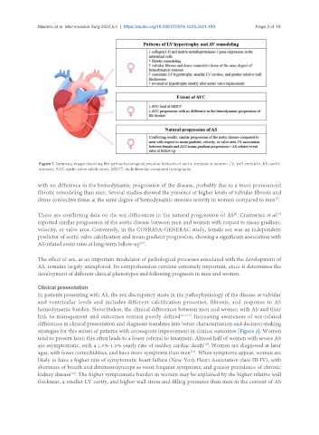
Figure 1. Summary image depicting the pathophysiological peculiar features of aortic stenosis in women. LV: Left ventricle; AS: aortic
stenosis; AVC: aortic valve calcification; MDCT: multidetector computed tomography.
with no difference in the hemodynamic progression of the disease, probably due to a more pronounced
fibrotic remodeling than men. Several studies showed the presence of higher levels of valvular fibrosis and
dense connective tissue at the same degree of hemodynamic stenosis severity in women compared to men .
[7]
[9]
There are conflicting data on the sex differences in the natural progression of AS . Cramariuc et al.
[8]
reported similar progression of the aortic disease between men and women with respect to mean gradient,
velocity, or valve area. Conversely, in the COFRASA-GENERAC study, female sex was an independent
predictor of aortic valve calcification and mean gradient progression, showing a significant association with
[10]
AS-related event rates at long-term follow-up .
The effect of sex, as an important modulator of pathological processes associated with the development of
AS, remains largely unexplored. Its comprehension remains extremely important, since it determines the
development of different clinical phenotypes and following prognosis in men and women.
Clinical presentation
In patients presenting with AS, the sex discrepancy starts in the pathophysiology of the disease at valvular
and ventricular levels and includes different calcification processes, fibrosis, and response to AS
hemodynamic burden. Nevertheless, the clinical differences between men and women with AS and their
link to management and outcomes remain poorly defined [7,11,12] . Increasing awareness of sex-related
differences in clinical presentation and diagnosis translates into better characterization and decision-making
strategies for this subset of patients with consequent improvement in clinical outcomes [Figure 2]. Women
tend to present later; this often leads to a lower referral to treatment. Almost half of women with severe AS
are asymptomatic, with a 1.0%-1.5% yearly rate of sudden cardiac death . Women are diagnosed at later
[13]
ages, with fewer comorbidities, and have more symptoms than men . When symptoms appear, women are
[12]
likely to have a higher rate of symptomatic heart-failure (New-York Heart Association class III-IV), with
shortness of breath and dizziness/syncope as most frequent symptoms, and greater prevalence of chronic
kidney disease . The higher symptomatic burden in women may be explained by the higher relative wall
[14]
thickness, a smaller LV cavity, and higher wall stress and filling pressures than men in the context of AS