
Page 70 - Read Online
P. 70
Page 4 of 8 Alam et al. Mini-invasive Surg 2021;5:48 https://dx.doi.org/10.20517/2574-1225.2021.65
Figure 1. Peritoneal breach showing underlying small bowel during dissection for a right inguinal TEP repair (A). Peritoneal breach
repaired by the use of 5 mm endo-clips (B). TEP: Totally extraperitoneal.
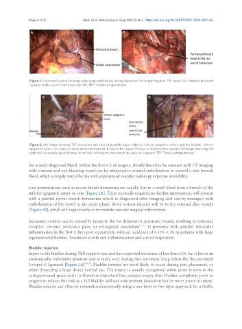
Figure 2. (A) Image showing TEP dissection and sites of possible injury with the inferior epigastric artery and the bladder. Inferior
epigastric artery can cause a rectus sheath hematoma. It should be clipped if injury is inadvertently caused. (B) Image depicting the
external iliac vessels, injury to these most likely will require intervention by vascular surgeons. TEP: Totally extraperitoneal.
An acutely diagnosed bleed, within the first 6 h of surgery, should therefore be assessed with CT imaging
with contrast and any bleeding vessel can be subjected to arterial embolization to control a side branch
bleed, which is largely very effective with experienced vascular radiology expertise availability.
Late presentations such as rectus sheath hematoma are usually due to a small bleed from a branch of the
inferior epigastric artery or vein [Figure 2A]. These normally required no further intervention, will present
with a painful rectus sheath hematoma which is diagnosed after imaging and can be managed with
embolization of the vessel in the acute phase. More serious injuries will be to the external iliac vessels
[Figure 2B], which will require early or immediate vascular surgical intervention.
Ischaemic orchitis can be caused by injury to the vas deferens or spermatic vessels, resulting in testicular
atrophy, chronic testicular pain, or retrograde ejaculation [16,17] . It presents with painful testicular
inflammation in the first 5 days post-operatively, with an incidence of 0.05%-0.1% in patients with large
inguinoscrotal hernias. Treatment is with anti-inflammatories and scrotal suspension.
Bladder injuries
Injury to the bladder during TEP repair is rare and has a reported incidence of less than 0.5% but it lies in an
anatomically vulnerable position and is easily seen during this operation lying below the ilio-pectineal
(cooper’s) ligament [Figure 2A] [18,19] . Bladder injuries are most likely to occur during port placement, or
when dissecting a large direct hernial sac. The injury is usually recognised when urine is seen in the
extraperitoneal space and it is therefore important that patients empty their bladder completely prior to
surgery to reduce this risk as a full bladder will not only prevent dissection but be more prone to injury.
Bladder injuries can often be repaired endoscopically using a one-layer or two-layer approach for a visible